本研究在清醒猕猴上颞沟(superior temporal sulcus, STS)下岸——中央下颞皮层(IT)后部 TE 区——开展单细胞记录,考察物体表面光泽(surface gloss)属性的神经表征。作者利用 MERL BRDF 数据集中 33 种实测材质的双向反射分布函数(BRDF)渲染物体图像,系统调节镜面反射率、漫反射率和粗糙度三个物理参数,筛选出一批对特定光泽选择性响应的神经元,并证实其选择性在改变物体形状或照明环境时依然保持,而在像素空间打乱后消失,展示了材质知觉在单细胞与群体编码层面的组织特征。
研究背景
物体识别不仅依赖轮廓形状和颜色,表面反射特性同样提供了关键的材质与物理状态信息。光泽感知对视觉系统提出了特殊的计算挑战:高光与明暗分布随物体三维形状及环境照明实时变化,但观察者仍能在不同图像间稳定感知到一致的表面光泽属性。在下颞皮层中,既往研究已发现面孔、纹理与颜色选择性细胞,上颞沟内部也记录到了与三维立体形状及光照方向相关的神经响应;人类功能磁共振成像与非人灵长类光泽—哑光对比研究进一步提示该脑区参与材质加工。然而,某皮层区域对光泽与哑光刺激表现出不同程度的激活,尚不能直接证明局部神经元提取的是表面反射特性,还是伴随表面变化而改变的对比度、局部亮度或几何线索。
此前针对材质选择性的单细胞电生理研究多采用真实材质摄影图像。这种刺激保留了自然外观,但也带来了特征归因上的困难:自然材质通常伴随中尺度微观表面起伏,由此产生的复杂明暗阴影与材料本身的反射特性交织在一起。单个神经元对高光表面的偏好,可能仅是对局部高亮度、特定色彩或某种局部形状的附带响应。要确证神经选择性直接对应表面光泽,需要引入互补的实验对照:一方面,在改变宏观物体形状导致局部亮度分布重组时,神经元对材质反射的偏好模式应保持稳定;另一方面,在维持全局亮度与色彩直方图但打乱空间结构破坏光泽知觉时,原有的选择性应当消失。此外,还需阐明这些细胞究竟仅是共同编码单调的明暗强度,还是能够通过群体反应模式区分多维度的表面反射特性。
本文利用实测反射数据渲染无宏观纹理起伏的三维几何体,在严格控制物体表面微观结构的条件下,系统调整漫反射率、镜面反射率与粗糙度参数,记录猕猴上颞沟下岸神经元的放电活动。研究通过更换物体形状、像素打乱以及切换自然照明环境,系统检验神经选择性的特征特异性与环境不变性,并通过群体反应相关距离分析构建神经表征几何构型。研究将光泽加工推进到了具有明确操作定义的单细胞与群体编码层面。需要说明的是,像素打乱在破坏高光的同时改变了多种高阶空间统计量,不能彻底排除所有高阶纹理特征假设;筛选标准也可能对低反射端的弱光泽细胞存在一定偏倚;群体距离与人类心理物理空间的吻合提示了光泽编码机制,但仍需结合行为学干扰研究进一步明确因果关系。
研究思路
研究设计了物理参数精准可控且贴近真实物理反射规律的刺激集:基于 MERL BRDF 数据集(包含约 100 种实测材料),选取 33 种表面反射特性,利用 Ward–Duer 反射模型沿漫反射率(ρd)、镜面反射率(ρs)与粗糙度(α)三个维度进行渲染,投射至 10 种无表面纹理起伏的三维模型上,默认采用桉树林(Eucalyptus Grove)高动态范围环境照明。由于渲染模型不存在微观凹凸纹理,排除了表面起伏阴影的干扰,使神经反应差异可归因于反射特性本身。
实验逻辑分为三步:第一,筛选出对特定表面反射展现出选择性反应的候选神经元;第二,实施两项互补对照以排除低级图像统计解释——更换三维几何形状(局部明暗分布彻底改变但光泽知觉保持不变,选择性模式应予以维持)以及轮廓内像素随机打乱(色彩与亮度直方图保持一致但空间高光结构瓦解、光泽知觉消失,选择性应随之消失),严格满足此两项标准的单位定义为光泽选择性(gloss-selective)神经元;第三,计算光泽选择性神经元群体反应向量的相关距离,应用多维标度分析(multidimensional scaling, MDS)探查光泽属性在神经群体活动中的空间表征结构。
方法
实验使用两只日本猕猴(Macaca fuscata,代号 AQ 与 TV,一雄一雌,体重 5.8–6.2 kg),在三个大脑半球开展记录。动物手术植入头部固定架与记录脑舱,先使用钨丝微电极在 STS 下岸(后部 TE 区,立体定位坐标 A4–A16、L18–L26)进行广泛电生理扫查,随后在稳定记录到光泽反应的区域植入核磁共振(MRI)兼容的金属引导管(钛/金合金),所有电极推进轨迹均经结构 MRI 扫描定位验证。记录采用被动注视任务:猕猴注视中央点后,以 300 ms 呈现、300 ms 间隔的时序连续呈现 5 幅图像,顺利维持注视在 2.6°×2.6° 窗口内至刺激结束后获得果汁奖励;刺激图像视角约为 5°,默认呈现于注视中心(反应微弱时移至感受野外周,共 27/215 个神经元、6/57 个光泽选择性神经元进行此项调整)。
数据分析中,神经元反应量取刺激呈现后 50–350 ms 窗口内的平均放电率并扣除刺激前基线,纳入标准为放电率 ≥10 spikes/s 且对至少 1 种刺激引出显著反应(ANOVA,p≤0.05)。实验先通过预实验(15 种反射特性 × 10 种形状)确定每个神经元的最优响应形状,随后在最优形状下测试 33 种反射特性。计算选择性指数(selectivity index = 1 − 最小反应/最大反应)与稀疏性指数(sparseness index,衡量放电在刺激集上的分布聚集度)。更换形状对照通过计算最优与非最优形状反应向量的 Pearson 相关系数及双因素 ANOVA 评估;像素打乱对照在物体轮廓内随机置换像素位置。光泽选择性神经元需同时满足两项判定标准:(1) 对非最优形状具有显著反应,且两种形状下的刺激选择性曲线呈现显著正相关(p≤0.05);(2) 对打乱刺激无显著反应,或打乱刺激下的选择性模式与原刺激不相关。另计算形状可分离性指数(separability index,基于奇异值分解,以第一主成分重构解释方差比 r² 评估光泽与形状因子的独立性),并结合 1000 次置换检验(permutation test)评估显著性。群体表征分析基于 57 个光泽选择性神经元对全部刺激对的反应相关系数(以 1−r 作为神经距离),执行非度量 MDS 分析。
主要结果
- STS 下岸存在对特定光泽展现特异性调谐的神经元。 在扫查的 101 个记录轨迹中,记录到 215 个对光泽刺激集有显著反应的神经元,其中 194 个表现出反射特性选择性;经严格双标准筛选,最终确立 57 个光泽选择性神经元(AQ 猴 31 个,TV 猴 26 个)。神经元呈现出差异化的表面偏好:示例 cell 1 专门响应具有锐利高光的表面(大 ρs、小 α),cell 2 偏好伴有弥散高光的平滑亮面(大 ρs、大 α),cell 3 则特异响应无明显高光的哑光表面(图 4)。
- 选择性独立于物体几何形状,由光泽属性主导。 更换三维形状后,神经元对 33 种反射特性的反应轮廓保持高度一致(cell 1 r=0.86,cell 2 r=0.82,cell 3 r=0.87;群体中 118/145 即 81% 的测试单位表现出显著相关,平均可分离性指数达 0.86±0.08,仅 1 个神经元的第二主成分显著);而轮廓内像素打乱后神经反应普遍丧失(cell 1 最大放电率仅 1.71 spikes/s,群体排序曲线趋近水平)(图 5、6、7、9)。
- 光泽选择性呈现尖锐调谐与稳健的统计特征。 57 个光泽选择性神经元的选择性指数中位数为 1.02(大部分 ≥0.6),稀疏性指数中位数为 0.43(大部分 ≥0.3);在最优形状与非最优形状下的选择性强度无统计学差异(1.03±0.18 对比 1.02±0.18);在最优与最差刺激条件下的微眼动方差仅在 1 个神经元上检出差异,排除了眼动对选择性的干扰(图 8)。
- 神经元反应偏好覆盖不同反射维度,群体放电偏向强光泽表面。 57 个神经元的归一化群体平均放电在最强刺激(刺激 13)与最弱刺激(刺激 33)间呈现 2.18 倍的动态范围,高反射率刺激整体引发更强放电;细胞的最佳偏好峰值多聚集于高镜面反射率(ρs)与低粗糙度(α)区域,低镜面反射表面较少引发峰值反应(图 10)。
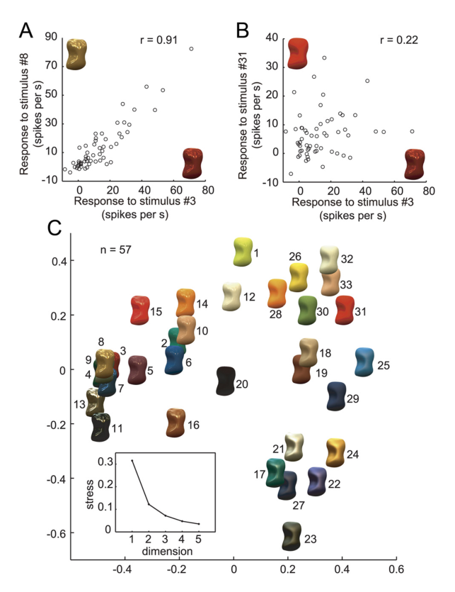
- 选择性跨越自然照明变化保持稳定,群体活动系统映射二维知觉空间。 在黄昏校园(Campus at Sunset)照明测试中,48 个受测单位中 40 个(83.3%)与默认光照下的选择性模式高度相关,平均照明可分离性指数达 0.84±0.1(仅 2 个神经元第二主成分显著)(图 11、12);MDS 二维分析能够充分拟合神经群体距离(stress 值为 0.12),高镜面反射表面聚集于左侧,模糊高光表面分布于右下方,哑光表面位于右上方,该神经构型与 Ferwerda 等(2001)提出的心理物理光泽空间维度(对比光泽 contrast gloss 与镜像清晰度 distinctness of image reflections)相吻合;外观相近的材质对(如 3 与 8)群体反应相关系数达 r=0.92,光泽迥异的材质对(如 3 与 31)相关系数仅为 r=0.22(图 13)。
图注解读
图 1 · 光泽三参数与刺激集构造
原文图注:Figure 1. Gloss parameters and stimuli for assessing gloss selectivity. A, Schematic illustration of three reflection parameters: diffuse reflectance (ρd), specular reflectance (ρs), and roughness (α). When ρd increases, the lightness of the object increases. When ρs increases, the highlights become stronger. When α increases, the highlights become blurred. B, Example of a gloss stimulus set. The stimuli exhibit 33 types of surface reflectance selected from the MERL BRDF dataset and rendered on one of the shapes (shape 3) under default illumination (Eucalyptus Grove). Stimuli were ordered according to the magnitude of α. C, Ten object shapes used for the experiment rendered with surface 8 in B. D, Top, Example of a shuffled stimulus generated by randomizing the pixels within the contour. Bottom, Example of a stimulus rendered under different illumination (Campus at Sunset). ……
A 展示基于 Ward–Duer 模型的三个物理反射参数:漫反射率(ρd)调控物体基础明度,镜面反射率(ρs)调节高光亮度,粗糙度(α)决定高光边缘的弥散模糊程度;B 展示采用默认桉树林环境光将 33 种表面反射渲染于几何形状 3 上的完整刺激集(按 α 递增排序);C 展示用于测试形状不变性的 10 种三维模型;D 分别展示轮廓内像素打乱刺激与备用照明(黄昏校园)刺激;E 则将 33 种表面投影在 ρd-ρs-α 构成的三维物理刺激空间中,建立了后续神经元调谐偏好的物理坐标系。
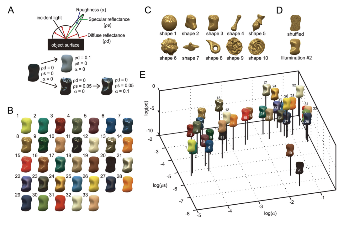
图 2 · 记录位点
原文图注:Figure 2. Recording sites. A, Schematic illustration showing the recording site within a lateral view of the monkey cerebral cortex (in red) and the approximate position of the recording chamber. B, An MRI image of a coronal section of the brain of monkey AQ positioned 8 mm anterior to the interaural line. A guide tube made of gold 650 μm in diameter is inserted targeting the lower bank of the STS in the right hemisphere. C, Top view of the areas of electrode penetration in the lower bank of the STS in three hemispheres are indicated by colored contours (red, monkey AQ right hemisphere; blue, AQ left; green, TV left) with stereotaxic coordinates. ……
A 在猕猴大脑侧面示意图上标示记录脑舱与上颞沟(STS)记录区域的大致解剖位置;B 展示猴 AQ 耳间线上方 8 mm 水平的冠状位结构 MRI 图像,金合金引导管精确穿向右半球 STS 下岸;C 在三个受测半球的记录俯视图中标出电极微导管穿透孔位,灰色圆圈代表对光泽刺激集有显著反应的记录位点,彩色实心点代表严格符合光泽选择性标准的神经元,明确了记录区域位于后部 TE 区的解剖边界。
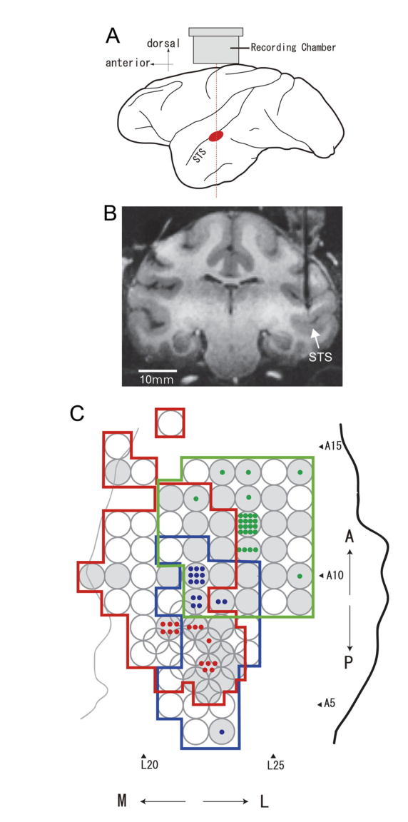
图 3 · 刺激示例:形状、打乱与照明
原文图注:Figure 3. Examples of stimuli. A, Examples of stimuli with 10 different shapes and 5 different surface reflectances rendered under default illumination (Eucalyptus Glove). B, Examples of shuffled stimuli (shape 3) with five different surface reflectances. C, Examples of stimuli (shape 3) with five different surface reflectances rendered under illumination 2 (Campus at Sunset).
A 展示 10 种形状与 5 种表面反射特性交叉组合的渲染效果,直观呈现了材质属性在不同几何载体间的视觉一致性;B 展示形状 3 在像素打乱条件下的对照图像,其色彩与亮度直方图保持不变,但宏观高光轮廓彻底解构;C 展示在黄昏自然照明环境下重新渲染的对应刺激,体现了光源变化导致的高光位移与色温重构,为后续不变性测试提供实验刺激范式。
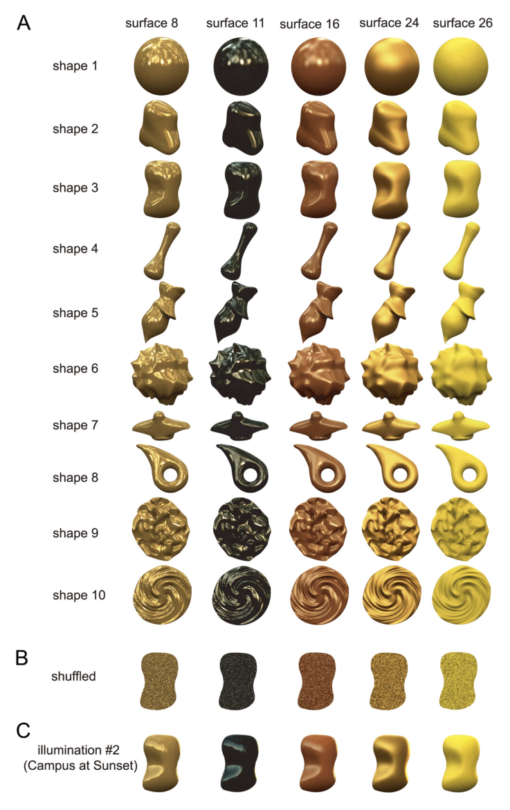
图 4 · 三个示例神经元:各有所好
原文图注:Figure 4. Responses to gloss stimulus set. A, Responses of an example neuron (cell 1) to the gloss stimulus set. The responses are depicted as raster plots and poststimulus time histograms (PSTHs). The horizontal bars under the PSTHs indicate the stimulus presentation period. B, Response magnitude of cell 1 to each stimulus in the gloss stimulus set represented by the size of the object image. This neuron strongly responded to stimuli with sharp highlights and did not respond to stimuli with weak glossiness. C, Response magnitude of cell 1 to each stimulus in the gloss stimulus set represented as the diameter of a circle and plotted at the corresponding position in the gloss stimulus space. D, E, Responses of another neuron (cell 2) ……. This neuron selectively responded to shiny objects with blurred highlights due to large specular reflectance and roughness. F, G, Responses of a third neuron (cell 3) ……. This neuron strongly responded to matte stimuli without clear highlights and to those with small specular reflectance and large roughness.
图中展示三个代表性神经元的电生理响应:A、B、C 展现 cell 1 的时间历程光栅图、PSTH 曲线,以及用刺激图标尺寸和三维反射空间圆圈直径表示的反应幅度,该神经元对锐利高光(小 α、大 ρs)产生高频放电;D、E 展现 cell 2 的响应特性,特异偏好伴有模糊边缘高光的光泽表面(大 α、大 ρs);F、G 展现 cell 3 的响应特性,对无高光的平光及弱光泽哑光表面呈现高放电。结果表明 STS 下岸神经元能够覆盖表面反射参数空间的不同子区域。
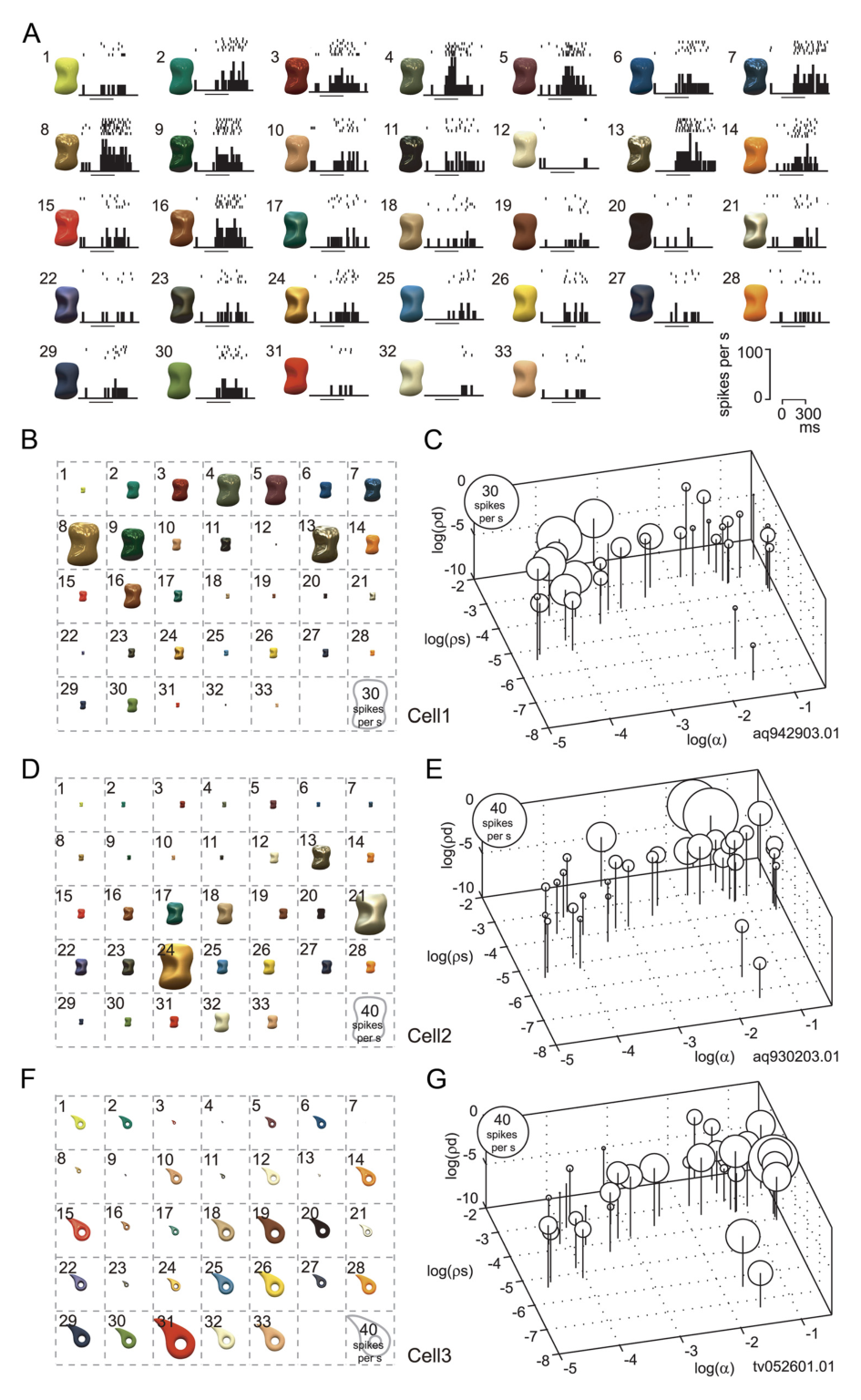
图 5 · 换形状与打乱像素:单细胞对照
原文图注:Figure 5. Effects of a change in object shape and pixel shuffling on the activity of the neurons depicted in Figure 4. A, Responses of cell 1 (the same neuron depicted in Fig. 4A–C) sorted according to the rank order of its response magnitude when the optimal shape was used. The horizontal axis indicates rank order for the optimal shape (shape 3); the vertical axis indicates response magnitude (with SEM). The red line depicts the responses to the optimal shape; the blue line, those to the nonoptimal shape (shape 2); and the black line, those to the shuffled stimuli. Object images with the optimal shape are shown at the top in rank order. The inset shows the relationship between the responses to each stimulus in the gloss stimulus set for the optimal (horizontal axis) and nonoptimal (vertical axis) shapes. B, C, Responses of cells 2 and 3 respectively, with the optimal and nonoptimal shapes 3/9 for cell 2 and 8/4 for cell 3.
以最优形状下的反应强度排序作为横坐标基准:cell 1 与 cell 2 在非最优形状下的放电曲线(蓝线)呈现同步单调下降,与最优形状(红线)高度相关(r=0.86、0.82),而像素打乱条件下的放电(黑线)几乎完全被抑制,证明其选择性依赖连续空间高光而非图像像素统计;相比之下,cell 3 虽然在换形状时表现出高相关(r=0.87),但在打乱像素后仍保留高幅度放电(黑线峰值达 25.6 spikes/s),提示其响应可能主要由低阶亮度或颜色分布驱动,因而被排除在光泽选择性群体之外。
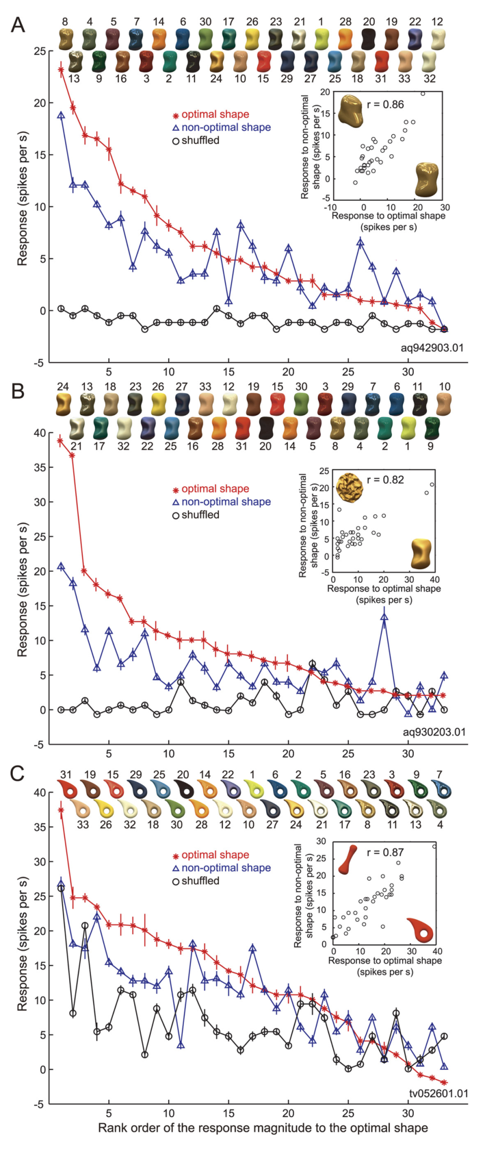
图 6 · gloss-selective 神经元的判定散点
原文图注:Figure 6. Effects of shape change and pixel shuffling: population analysis. In the scatter plot, horizontal axis indicates correlation coefficient between the responses to the optimal and nonoptimal shapes, and vertical axis that between the responses to the optimal shape and shuffled stimuli. If a neuron did not exhibit significant response to the nonoptimal shape or shuffled stimuli, they are plotted on the horizontal or vertical axis, respectively. We defined "gloss-selective" neurons using two criteria: (1) They should be responsive to a nonoptimal shape, and there should be significant correlation between the patterns of stimulus selectivity between the optimal and nonoptimal shapes (p≤0.05). And (2) they should not show significant response to shuffled stimuli ……. The red circles represent gloss-selective neurons that satisfied these two criteria. ……
散点图横轴为最优与非最优形状反应向量的相关系数,纵轴为最优形状与像素打乱刺激反应向量的相关系数。位于右下区域由红色圆圈标出的 57 个神经元同时满足跨形状调谐显著相关且对打乱刺激无响应或不相关两项标准,正式判定为光泽选择性神经元;蓝色圆圈标出的 43 个单位虽具备跨形状相关性,但在像素打乱后仍保留显著相关,提示其可能由低阶图像色彩或亮度直方图特征所驱动。
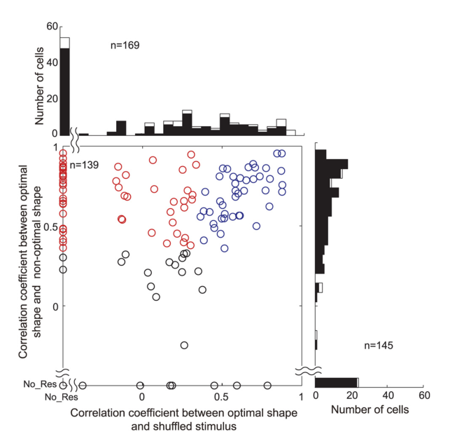
图 7 · 形状可分离性指数
原文图注:Figure 7. Separability index for a change in shape. A, Distribution of the separability index for a change in object shape. The horizontal axis indicates the separability index, the vertical axis the number of cells. B, Distribution of r² between the actual and predicted responses computed from only the second principal component. The filled and open bars indicate significant and nonsignificant cells, respectively, based on the permutation test. C, Raw interaction plot connecting the responses to the gloss stimulus set for the optimal shape (left) and the nonoptimal shape (right) for four representative example neurons. The left two panels are for cell 1 and cell 2 depicted in Figures 4 and 5. Separability index for each neuron is 0.92, 0.93, 0.96, and 0.98, respectively.
A 展现 57 个光泽选择性神经元的形状可分离性指数分布,数值集中在较高区间(平均 0.86±0.08,多数 ≥0.7),表明神经元对光泽刺激的调谐曲线在不同形状下主要表现为幅度线性增益变化;B 显示基于置换检验计算的第二主成分解释方差,仅 1 个单位达到显著水平,说明光泽与形状因子的非线性交互作用微弱;C 展示 4 个代表性细胞的放电交互线段图,连接最优与非最优形状的反应线段呈现高度平行走势。
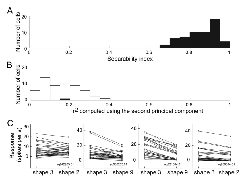
图 8 · 选择性与稀疏性分布
原文图注:Figure 8. Distribution of the selectivity and sparseness indices among gloss-selective neurons. A, Distribution of the selectivity indices of 57 gloss-selective neurons. The horizontal axis indicates the selectivity index, and the height of each bar indicates the number of cells (left vertical axis). The rightmost bar indicates cells with a selectivity index over 1.2. The black line indicates the cumulative percentage of indices (right vertical axis). B, Distribution of the sparseness indices of the 57 gloss-selective neurons. Other conventions are the same as in A.
A 展示 57 个光泽选择性神经元的选择性指数频数直方图与累积百分比曲线,中位数为 1.02,大多数神经元数值大于 0.6,反映出高对比的选择性响应;B 展示稀疏性指数分布,中位数为 0.43,表明各神经元仅对测试集中特定范围的反射参数产生集中放电,呈现出针对特定材质属性的尖锐调谐特性。
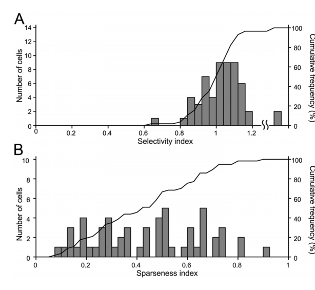
图 9 · 群体平均的排序曲线
原文图注:Figure 9. Rank order of the responses to the gloss stimulus set: population average. A, Average of the responses of 57 gloss-selective neurons (Fig. 6, red circles) to stimuli with the optimal shape (red line), a nonoptimal shape (blue line), and shuffled stimuli (black line), sorted according to the rank order of the responses to the optimal shape for each neuron. B, Average of the responses of 43 neurons that showed significant correlation between the responses to the optimal shape and shuffled stimuli (Fig. 6, blue circles). Other conventions are the same as in A.
A 中 57 个光泽选择性神经元的群体平均放电在非最优形状(蓝线)下随最优形状排序(红线)保持清晰的单调递减趋势,而打乱刺激(黑线)的平均反应近乎平缓贴近基线;B 中 43 个疑似低阶特征驱动单位在打乱刺激下的群体放电依然随原排序呈现显著递减,直观展示了筛选标准在区分真性光泽编码与低阶图像统计特征中的有效性。
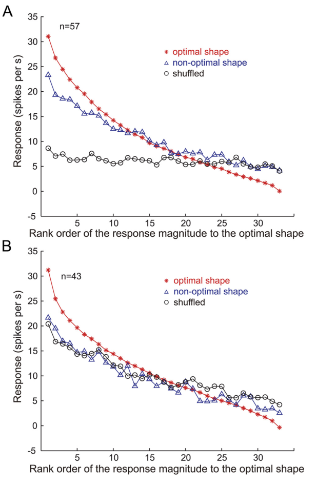
图 10 · 偏好刺激的分布
原文图注:Figure 10. Stimulus preference of gloss-selective neurons. A, C, Responses of a gloss-selective neuron that was selectively responsive to shiny objects with clear highlights (cell 4). B, D, Responses of another gloss-selective neuron that was selectively responsive to matte objects (cell 5). These are examples of gloss-selective neurons recorded from monkey TV, while those shown in Figure 4 (cell 1 and cell 2) are from monkey AQ. Conventions are the same as in Figure 4, B and C. E, Population average of the normalized responses of 57 gloss-selective neurons to each stimulus in the gloss stimulus set. F, The numbers of gloss-selective neurons that showed a peak response to each stimulus in the gloss stimulus set.
A–D 展示第二只猕猴(TV)记录到的极端偏好细胞示例(强高光选择性 cell 4 与哑光选择性 cell 5),确认不同反射偏好在个体间具有普遍性;E 展示 57 个神经元归一化平均放电率对 33 种刺激的响应谱,整体呈现出对高镜面反射刺激的群体响应偏向;F 统计了以各刺激作为最大响应峰值的细胞数量分布,峰值细胞集中分布于大 ρs 与低 α 区域,反映出群体表征在参数空间中的非均匀密度。
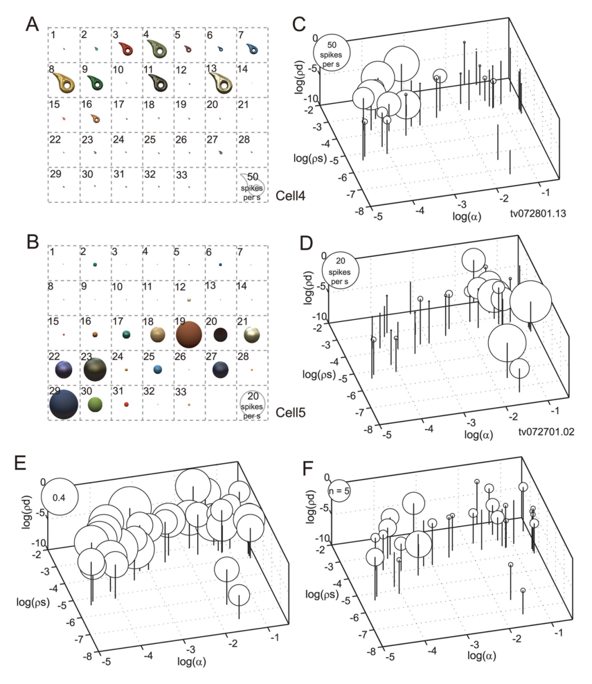
图 11 · 照明改变:单细胞与群体汇总
原文图注:Figure 11. Effects of illumination change. A, Responses of cell 1 (the neuron depicted in Fig. 4A–C) sorted according to the rank order of the response magnitudes under the default illumination (illumination 1, Eucalyptus Grove). The horizontal axis indicates the rank order of the responses, and the vertical axis indicates the response magnitude (with SEM). The red line depicts the responses under illumination 1; the blue line depicts those under different illumination (illumination 2, Campus at Sunset). ……. The inset shows the relationship between the responses to each stimulus in the gloss stimulus set under illuminations 1 (horizontal axis) and 2 (vertical axis). B, Summary of the effects of the illumination and shape in 48 gloss-selective neurons tested under both illuminations. ……
A 展示示例 cell 1 在切换为黄昏校园自然照明环境(蓝线)后,其放电强度沿默认桉树林照明下的排序基准仍保持显著递减(r=0.81);B 总结了 48 个在双照明下完成测试的神经元数据,其中 40 个神经元(83.3%)在不同照明环境间保持了显著正相关的刺激选择性模式,证实了光泽选择性在自然光照变化下的鲁棒性。
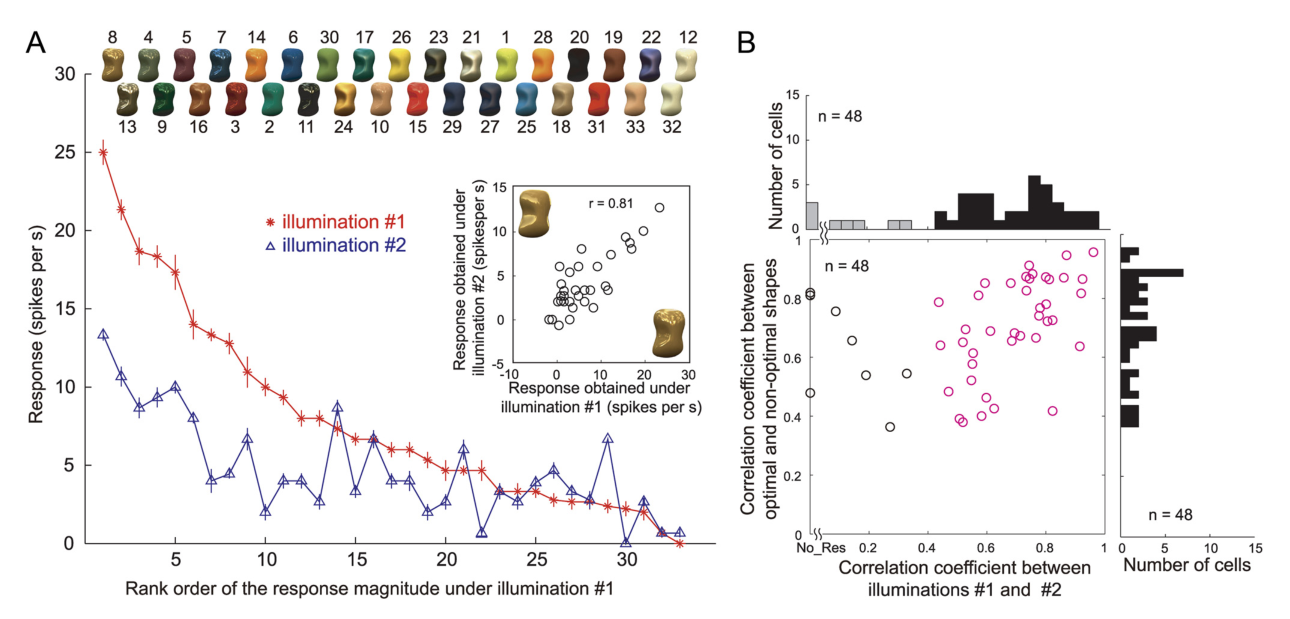
图 12 · 照明可分离性
原文图注:Figure 12. Effect of illumination on responses among gloss-selective neurons. A, Distribution of separability indices for the change in illumination. The filled and open bars indicate significant and nonsignificant cells, respectively, based on the permutation test. B, Distribution of r² between the actual and predicted responses computed from only the second principal component. C, Average of the responses of 57 gloss-selective neurons to stimuli with the optimal shape rendered under default illumination (illumination 1, Eucalyptus Grove; redline) and another illumination (illumination 2, Campus at Sunset; blueline) sorted according to the rank order of the responses under illumination 1.
A 显示照明可分离性指数分布,48 个神经元中除 1 个外均达到统计学显著(平均值 0.84±0.1);B 显示第二主成分解释方差仅在 2 个单位中达显著;C 展示 57 个神经元在两种照明下的群体排序响应均线,呈现出一致的平滑下降走势,表明照明维度的改变同样以整体尺度增益为主,未发生调谐特性的显著形变。
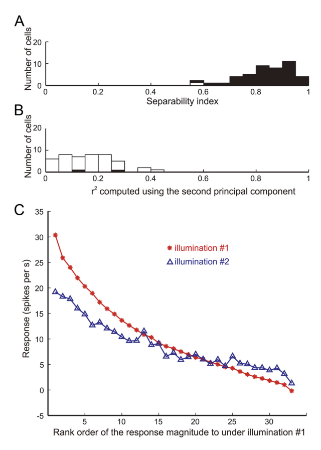
图 13 · 群体表征的 MDS 平面
原文图注:Figure 13. Neural representation of gloss in the activities of gloss-selective neurons. A, Relationship between the responses of 57 gloss-selective neurons to a pair of stimuli (surfaces 3 and 8) that are similarly glossy in appearance. The horizontal axis indicates responses to one stimulus (no. 3); the vertical axis indicates the responses to the other (no. 8). B, Relationship between the responses of 57 gloss-selective neurons to a pair of stimuli (surfaces 3 and 31) that differ with respect to their glossiness. Conventions are as in A. C, Two-dimensional plot of the results of MDS analysis. Distances were based on 1 − r between the responses of the 57 gloss-selective neurons for each stimulus pair from the gloss stimulus set. The inset is a scree plot showing the relationship between the number of dimensions and the stress in the MDS analysis.
A 与 B 通过代表性刺激对展现群体反应向量距离:外观相近的高光表面对(3 与 8)群体反应向量高度线性相关(r=0.92),而光泽迥异的刺激对(3 与 31)相关系数仅为 0.22;C 展示基于群体相关距离(1−r)的二维 MDS 降维拓扑图(内嵌碎石图显示二维应力值 stress 为 0.12),测试材质在平面内按光泽物理特性呈现连续排布,并在拓扑结构上对应于人类心理物理学中的对比光泽与高光清晰度维度。
讨论
作者结合实验发现对下颞叶皮层的材质加工机制进行了系统探讨。首先,这批神经元表现出的跨形状不变性、跨自然照明不变性以及对像素空间重排的高度敏感性,表明其放电活动对应于与物体表面反射属性直接绑定的视觉表征,而非局限于特定位置的低级几何轮廓或图像统计直方图。由于高光与明暗模式随光源和观察角度动态移动,而表面纹理与双目视差线索则相对固定于物体表面物理坐标系,作者推测在 STS 下岸区域可能存在功能分离的并行通路:一部分皮层机制利用双目视差与纹理梯度表征三维几何结构(如 Janssen 等报告的更偏前部的三维形状选择区),另一部分机制则专门整合明暗渐变与高光线索以抽取表面反射属性;同时,该记录位点也与已知的色彩选择性集中区保持解剖分离。
其次,在方法学层面,相较于既往直接使用自然材质摄影图像的研究,实测 BRDF 渲染去除了材质自带的微观表面起伏,将纯粹的表面物理反射特性与宏观几何阴影进行了有效解离,使对神经调谐特异性的归因更为明确。第三,群体放电构建的神经拓扑空间与人类知觉光泽空间维度的吻合,表明 IT 皮层的群体活动能够为材质知觉提供结构化的表征基础;但作者同时指出,视觉皮层具体通过何种中间图像特征计算并生成此种选择性,仍有待未来进一步阐明。在分类边界上,光泽选择性神经元与更广泛的视皮层神经元之间呈现连续谱系分布,筛选标准较为保守,可能未能完全覆盖偏好低反射率或极端哑光表面的神经元群。总体而言,研究确立了灵长类下颞皮层在物体表面反射与光泽感知加工中的关键地位。
一句话总结
研究通过变换几何形状保留反射属性与打乱像素消除高光结构的互补实验,证实下颞叶神经元具备独立于几何形状与照明条件的光泽选择性,其群体反应构型与知觉光泽空间维度相吻合,揭示了高级视觉皮层对物体表面材质属性的结构化表征。