本综述探讨了短波敏感视锥(S 视锥)信号在背侧与腹侧视觉通路中的功能分化,重点对比了中颞区(MT)与 V4/后下颞叶皮层(PIT)。文章区分了神经元的“颜色敏感性”与“颜色选择性”,并提出双重解释:在腹侧通路的颜色特化区域,S 视锥输入参与将视锥拮抗信号转化为知觉色调表征;而在背侧通路(如 MT)及腹侧非颜色偏好区域中,S 信号主要提供与环境光照相关的线索(如日光照耀下的黄与阴影处的蓝),支持从阴影中提取三维空间结构与运动信息。
研究背景
色觉研究传统上从视网膜 L、M、S 三类视锥的光谱吸收出发,经由外侧膝状体(LGN)追踪至初级视皮层(V1)。然而,LGN 神经元的色度拮抗轴主要对应于“红-青”与“薰衣草-青柠”等互补轴,并不等同于主观感知的对立色;部分细胞的空间拮抗模式亦与色觉对比计算的预期存在差异。S 视锥在光感受器中所占比例较低,但能显著激活 V1 细胞,且其输入的空间符号常与 M 视锥输入对齐。因此,从外周视锥拮抗信号向皮层知觉色调的转换机制仍需阐明。
在纹外皮层中,背侧通路(如 MT)主要参与运动与空间视觉加工,腹侧通路(如 V4 及下颞叶)主要处理物体形状与表面属性。然而,电生理与神经影像研究表明,S 视锥刺激不仅能激活腹侧颜色偏好区,同样能驱动 MT 神经元。若仅依据对色度刺激产生电位改变即推断其参与色彩感知,会导致功能定位的泛化。为此,作者强调区分“颜色敏感性”(神经元对色度变化存在反应)与“颜色选择性”(神经元具备亮度不变的稳定色调调谐)。这使得检验 S 信号在不同通路中的特异功能成为可能。
基于此,作者提出:在腹侧通路的颜色偏好区,S 视锥输入用于扩展知觉色轮的蓝色维度;而在背侧通路或非颜色偏好区,S 信号更可能提供与环境光照及阴影相关的低频空间线索。
研究思路
文章结合通路层级追踪与跨区域比较:纵向比较 LGN、V1、MT 以及 PIT 内部颜色偏好区与非偏好区对相同颜色刺激(定义于 DKL 视锥拮抗空间等亮度平面的光栅)的神经响应;横向整合单细胞微电极记录、逆行神经解剖示踪以及猕猴与人类的功能磁共振成像(fMRI)数据。
分析重点考察两套编码转换模型:从皮层下的群体编码(以 L-M 与 S-(L+M) 两主轴加权平均)向皮层的区间编码(尖锐色调调谐神经元集合)的演变。针对 S 视锥信号的功能,研究提出双重定位:在 PIT 颜色特化区(globs),S 输入用于构建均衡的二维色相空间;在非颜色特化区(MT 及 PIT 间隔区),S 信号通过反映自然光照的色彩梯度(直射阳光偏黄、天光阴影偏蓝),辅助基于阴影的场景三维结构感知与运动分析。
方法
综述引用的实证数据包含作者团队在清醒猕猴上完成的 fMRI 实验(Lafer-Sousa et al. 2012)。刺激采用异色光栅(空间频率 2.9 周/度,漂移速度 0.75 周/秒,每 2 秒反转运动方向),对应 DKL 等亮度平面上的四个色轴:红-绿(L-M 轴)、蓝-黄(S 轴)、橙-青(日光轴)与青柠-品红(正交中间轴)。两组中间轴对底层 L-M 与 S 主轴机制的激活总量相匹配,以排除由底层视锥对比度差异造成的激活偏差。
电生理记录主要涵盖清醒猕猴的单单位微电极实验:在 V1 中通过稀疏噪声视锥分离刺激结合反相关技术重构双对立感受野;在 PIT 中利用 fMRI 引导微电极针对 glob 与 inter-glob 区域进行记录,并结合不同背景明度刺激检验色调调谐的亮度不变性。
主要结果
- MT 区具备颜色敏感性但缺乏色调选择性:MT 神经元对等亮度运动刺激的放电率通常低于含亮度对比的刺激,但在缺少亮度线索时,等亮度色度刺激仍能有效驱动 MT 神经元;在红-蓝光栅的等亮度测试中,MT 的 BOLD 响应在主观等亮度点降低但未消失。然而微电极记录未发现具备亮度不变性的色调选择性 MT 神经元,说明 MT 具备色度敏感性而非用于知觉色调分类。
- S 视锥输入进入 MT 的解剖与功能通路:清醒猕猴单细胞记录显示 MT 神经元对 S 视锥分离刺激存在响应;人类 fMRI 显示 MT 的 S 刺激响应幅度约为 V1 的 2 倍(亮度响应约为 6 倍)(图 3)。逆行示踪研究表明,LGN 粒状细胞层中存在直接投射至 MT 且绕过 V1 的神经元(图 4),构成了 S 信号快速输入背侧通路的解剖学基础。
- 皮层加工中日光轴响应偏置的出现(图 5、6):LGN 细胞群体的调谐峰对齐于 L-M 与 S 轴,未见显著的日光轴偏置;在 V1 中,群体 fMRI 响应沿橙-青(日光轴)的反应幅度显著高于正交的青柠-品红轴(图 5);鉴于两中间轴对底层主轴机制激活强度一致,该差异反映了 V1 内部对主轴输入的非线性组合。MT 及 PIT 的非偏色间隔区(PITi)同样表现出日光轴偏置(图 6B、C)。
- PIT 颜色偏好区对 S 轴信号的增益与坐标转换(图 6D、8、10):PIT glob 细胞对视锥分离刺激的反应依赖于刺激与细胞最适色调的匹配(图 8);绿色调谐神经元对 M+、L-、S- 产生的绿色刺激均有响应,与 V1 拮抗细胞的输入极性不同,反映了由视锥拮抗空间向知觉色调空间的转换。PIT 颜色偏好区(PITc)相对 V1 表现出针对 S 轴响应的特异性增强,促进了色轮各区域表征幅度的均衡化。此外,初步测量提示 glob 细胞调谐对饱和度变化保持稳定,且较低饱和度伴随更长的放电潜伏期(图 10)。
- PIT 颜色敏感神经元的拓扑聚集(图 9):沿微电极记录轨迹,相邻 glob 神经元的最适色调呈现按色轮顺序(红-橙-紫等)的连续梯度分布,形成色位图(chromatopic maps);偏离局部梯度的少数神经元表现出相对延后的反应潜伏期。
- 非颜色区 S 信号的场景分析功能(图 7):S 视锥在视网膜中心凹分布稀疏且受眼屈光色差限制,其解剖与光学特性天然偏向低空间频率加工。在自然日光环境中,受直射日光照射区域偏黄橙色,而受天空散射光照明的阴影区域偏蓝;这一蓝-黄色度梯度能够提供关于三维表面形貌的信息,非颜色区对 S 信号的敏感性有助于在复杂光照下从阴影中解析场景结构与运动。
图注解读
图 1 · 背腹双通路示意图
原文图注:Figure 1. Simple schematic of the brain showing the visual pathway from the retina, through the dorsal lateral geniculate nucleus of the thalamus (LGN), up to primary visual cortex (V1). From V1, through intermediate visual areas (V2, V3, V4, not labeled), two major routes have been described, a dorsal pathway through MT (and related areas), and a ventral pathway through posterior, central and anterior inferior temporal (IT) cortex. The dorsal pathway has been implicated in encoding dynamic spatiotemporal relationships among visual objects (object action), while the ventral pathway is thought to represent stable attributes of objects (object quality)(DiCarlo et al., 2012; Kravitz et al., 2012). S-cone activation drives responses within both dorsal and ventral pathways. IT contains regions that are relatively more responsive to color (one such region is shown hatched in PIT).
展示视网膜经 LGN 到达 V1,再分支为经 MT 的背侧通路与经下颞叶的腹侧通路的示意图。标注了背侧通路处理运动时空关系、腹侧通路处理物体稳定属性的分工,并指明 S 视锥激活同时传入两条通路。
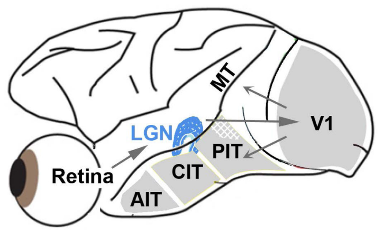
图 2 · V1 双拮抗细胞的锥体输入空间结构
原文图注:Figure 2. Spatial organization of cone inputs to a double-opponent cell recorded in alert macaque primary visual cortex. A, Spatial receptive-field map generated using sparse noise cone-isolating stimuli and reverse correlation. Scale of the small divisions on the grid is 0.75° of visual angle. The receptive-field center of the cell was activated by the "+" direction of the L-cone stimulus and the "-" direction of the M-cone stimulus, both of which appear reddish, but also by the "-" direction of the S stimulus, which appears lime. Note that the colors associated with the optimal polarity for each of the three cone-isolating stimuli do not fall in the same category, undermining the claim that the neuron contributes directly to hue. The inset gives an indication of the color of each stimulus (although the actual stimuli were presented on a computer monitor and carefully color calibrated). A "+" stimulus causes a selective increase in activation of the given cone type (compared to the activation generated by the adapting background); a "-" stimulus causes a selective decrease in cone activation. B, Spike-triggered average traces. The receptive-field center (central region in panels, A) was excited by an increase in L-cone activity (L+) or a decrease in M or S activity (M–, S–), and suppressed by a decrease in L (L–) or an increase in M or S (M+, S+); the receptive-field surround gave the opposite pattern of responses. Data from (Conway, 2001; Conway & Livingstone, 2006).
(A) 利用稀疏噪声视锥分离刺激重构的 V1 神经元二维感受野空间图:中心受 L+、M- 及 S- 激活,三者对应不同色相刺激;(B) 动作电位触发平均波形展示中心与外周的拮抗特性(中心受 L+、M-、S- 兴奋,受相反极性抑制;外周呈现相反极性)。表明该细胞执行空间色度对比计算而非直接对应单一感知色相。
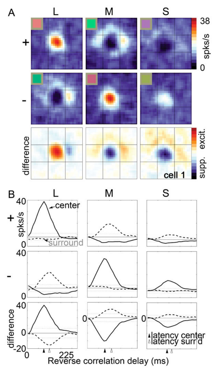
图 3 · 人类 fMRI:V1 与 MT 对亮度与 S 视锥刺激的响应
原文图注:Figure 3. Responses measured with fMRI in human subjects to stimuli with luminance contrast (squares) or S-cone contrast (diamonds). A, V1 responses. B, MT responses. The shape of the S-cone response function in V1 is not a scaled version of the luminance response, consistent with the conclusion that the response to the two stimuli are mediated by different underlying neural components (i.e. the S-cone responses is not just driving the luminance channel weakly). Luminance responses in MT are ∼6X greater than luminance responses in V1; S-cone responses in MT are ∼2X greater than those in V1. From (Wandell et al., 1999).
对比人类 V1 (A) 与 MT (B) 对亮度对比(方块)与 S 视锥对比(菱形)的 BOLD 反应函数。V1 内 S 视锥响应形态与亮度响应不同;MT 对两类刺激均有响应,且对亮度刺激的增益显著高于对 S 视锥刺激的增益。
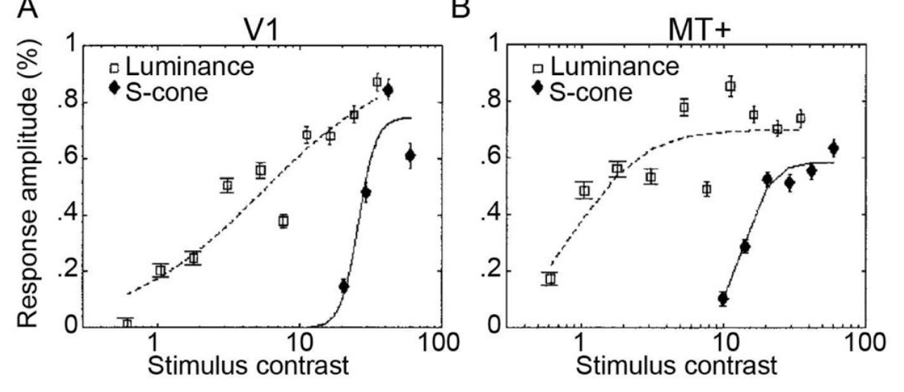
图 4 · LGN 粒状细胞层向 MT 的直接投射
原文图注:Figure 4. Neurons in macaque LGN identified after retrograde tracer injection in area MT and V1. A, Low-power of a slice through the entire LGN showing MT-projecting cells in red and V1-projecting cells in black. B, High-power of the boxed region in A. MT-projecting cells labeled in blue; V1-projecting cells labeled in brown. Note the two MT-projecting neurons that are conspicuously not double-labeled, showing that the axons of these neurons project directly to MT and bypass V1. MT-projecting neurons appear to be located preferentially within the koniocellular layers of the LGN, which are thought to carry strong S-cone signals. Data from (Sincich et al., 2004).
向猕猴 MT 与 V1 注射逆行示踪剂后的 LGN 组织切片显微图:(A) 低倍视野展示向 MT 投射的细胞(红色)与向 V1 投射的细胞(黑色);(B) 高倍视野显示单标蓝色的神经元直接投射至 MT 且绕过 V1,解剖上多位于富含 S 视锥信号的粒状细胞层。
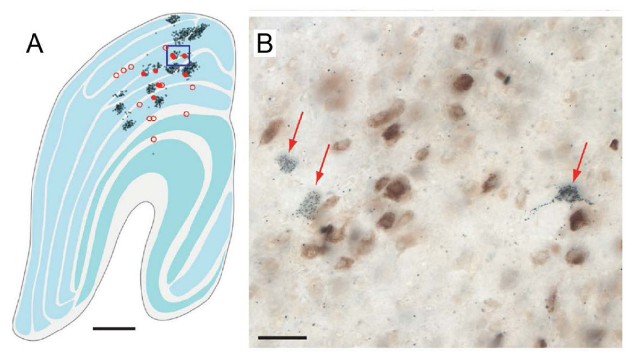
图 5 · V1 锥拮抗细胞群体对日光轴的偏置
原文图注:Figure 5. Cone-opponent cells in V1 are biased in chromatic tuning for colors of the daylight axis. A, responses of a population of cone-opponent neurons to cone-isolating stimuli, projected on the cone-opponent axes. Inset top right shows the standard C.I.E. chromaticity diagram with the cone-opponent axes intersecting at the neutral point, and the chromaticities of many samples of daylight. Inset top right shows the equiluminant plane through the DKL color space. Note that the 45° axis extending from –S/L-M through the origin towards +S/M-L forms the "daylight" axis. B, Macaque V1 responses measured using fMRI to different color directions of the DKL color space. Note the bias for the daylight axis. Responses have been normalized to 1. Stimuli comprised heterochromatic gratings, 2.9 cycles/degree, drifting slowly at 0.75 cycles/second, reversing direction every 2 seconds. Data from experiment 1 in (Lafer-Sousa et al., 2012).
(A) V1 视锥拮抗细胞群体对视锥分离刺激的响应投影,插图显示沿 −S/L-M 至 +S/M-L 分布的日光色度轴;(B) 猕猴 V1 对 DKL 空间各颜色轴的 fMRI 响应,显示沿日光轴的激活显著高于正交中间轴。
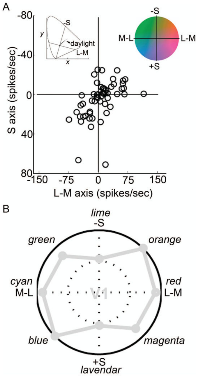
图 6 · 从 LGN 到 PIT 各阶段的色轴响应对比
原文图注:Figure 6. Responses measured using fMRI to different color directions of the DKL color space at multiple stages of visual processing. A, Responses within the LGN. B, Responses in MT. C, Responses in the portions of posterior IT that do not show a color bias ("i" for "in between" color-biased regions). D, Responses within color-biased regions of PIT. Responses in V1 shown in gray for comparison. Other conventions as for Figure 5B. PITi shows a pronounced bias along the daylight axis. Data from experiment 1 in (Lafer-Sousa et al., 2012). Cone contrasts for the four chromatic conditions given in Table 1 (adapted from Table 2 of Lafer-Sousa et al., 2012). Note that the two intermediate axes would elicit the same average activation of the underlying cardinal mechanisms, and that the total cone contrast (sum of L, M and S-cone contrasts) was much higher for the S stimulus than for the L-M stimulus. Despite the higher cone contrast, the response to the S stimulus was relatively low in LGN, V1, MT and PITi compared to the response to the L-M stimulus. PITc showed a relatively stronger response to S cones compared to V1. The increase in response along the S-axis within PIT color-biased regions achieves a more balanced response to all colors around the color wheel, and is consistent with the more likely involvement of this region in encoding color perception.
四个面板展示 LGN (A)、MT (B)、PIT 非偏色间隔区 PITi (C) 及 PIT 颜色偏好区 PITc (D) 对 DKL 色轴的响应幅度,灰色线为 V1 对照。结果表明仅在 PITc 区域观察到相对 S 轴响应的增强,促进了对全色相空间的均衡表征。
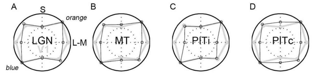
图 7 · 莫奈画中的蓝影子
原文图注:Figure 7. A, Morning Snow Effect (1891), oil on canvas (65.4×92.4cm), by Claude Monet. Museum of Fine Arts, Boston. B, Haystacks (Sunset)(1891), oil on canvas (73.3×92.7 cm), by Claude Monet. Museum of Fine Arts, Boston. Monet routinely depicts shadows with blue, surrounded by daylight yellow-orange.
莫奈油画作品展示:以蓝色调表现阴影、以黄橙色表现日照区域,直观说明自然场景中蓝-黄色度梯度与空间几何分布的对应关系。
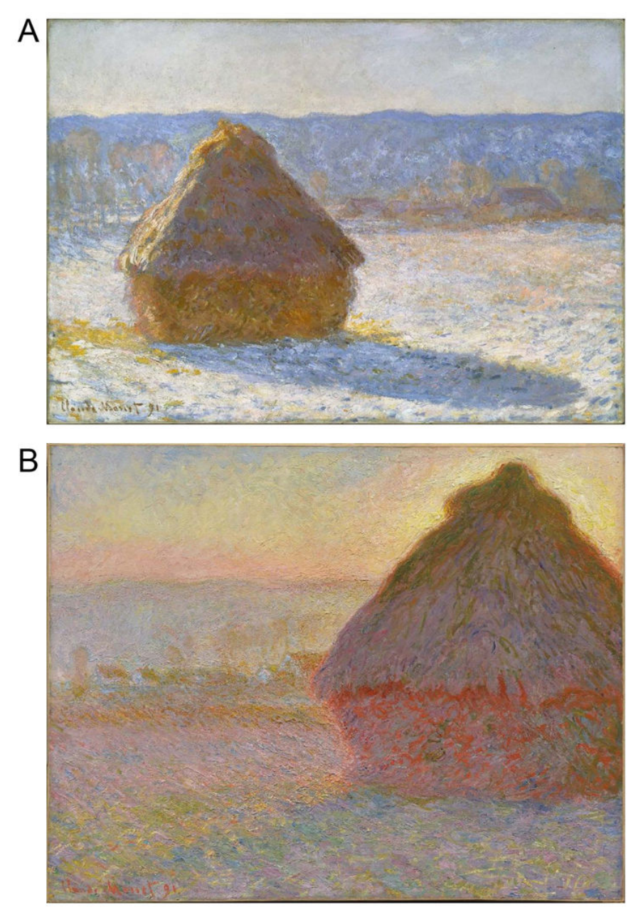
图 8 · PIT 绿色调 glob 细胞的朝向与颜色调谐
原文图注:Figure 8. Cone inputs to a green-tuned glob cell in PIT. A, Orientation tuning (max. 2 spikes/sec.) B, Color tuning (thick, medium and thin lines show tuning to higher, equal-with-background and lower luminance stimuli). C, L, M and S cone inputs. Compare with V1 cell (Fig. 2). Unpublished data of the author; see (Conway et al., 2007).
展示 PIT glob 绿色调谐神经元的生理响应:(A) 朝向调谐;(B) 在三种背景亮度下的颜色调谐曲线,显示调谐峰保持稳定;(C) 视锥输入极性显示其整合了导致绿色知觉的各类视锥输入组合。
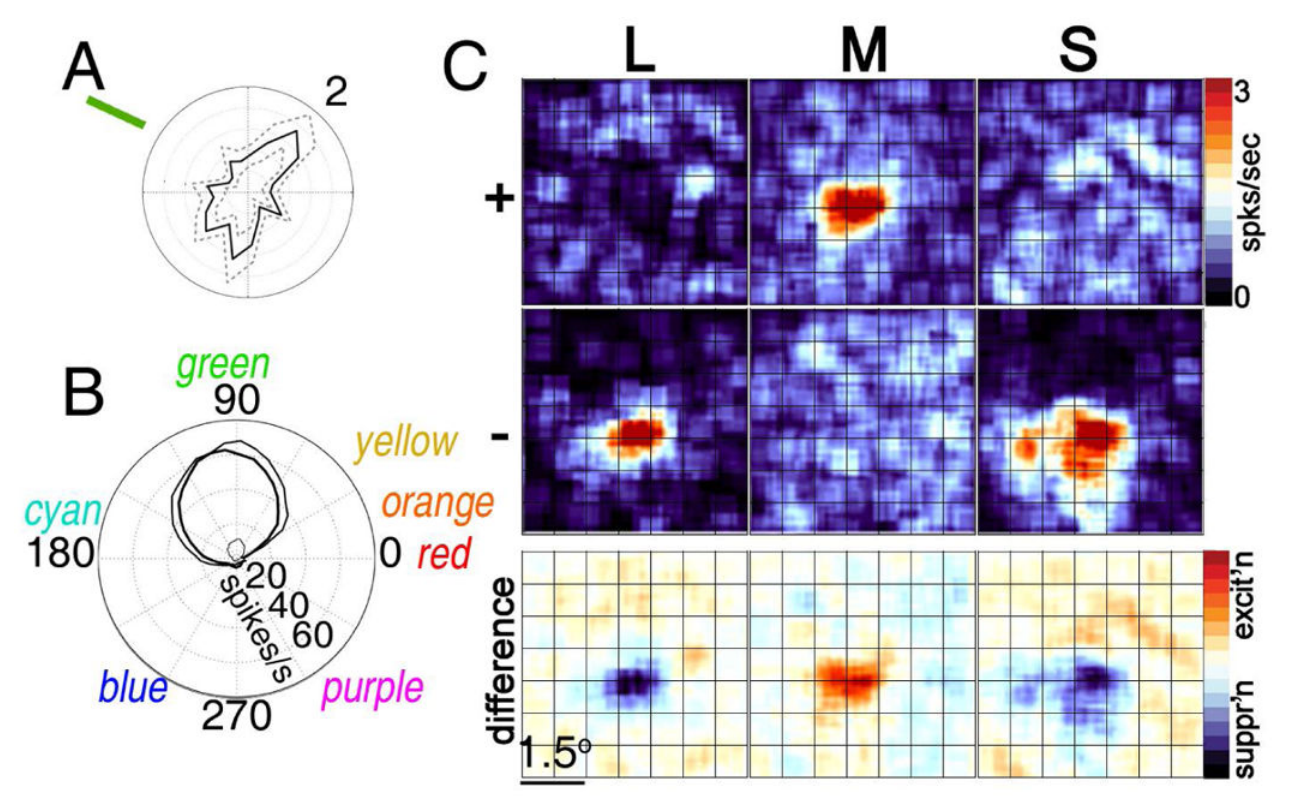
图 9 · PIT 颜色调谐细胞的"色位图"聚集
原文图注:Figure 9. Color-tuned neurons in PIT globs are clustered by color selectivity, and arranged according to "chromatopic" hue maps. A, Anatomical MRI of electrode targeting a color-biased region (glob). Scale, 1cm. B, Color tuning of six sequentially encountered neurons. Polar coordinates as for Figure 8B. C, Color tuning of all neurons encountered along the electrode path. Gray symbols show "outliers". D, Latency of the responses of the neurons. Data from (Conway & Tsao, 2009).
(A) 记录电极定位于 PIT glob 的结构 MRI;(B, C) 沿电极穿刺轨迹依次记录的神经元最适色调分布,呈现顺次渐变的色度图谱排列;(D) 各神经元的放电潜伏期分布,离群神经元潜伏期相对较长。
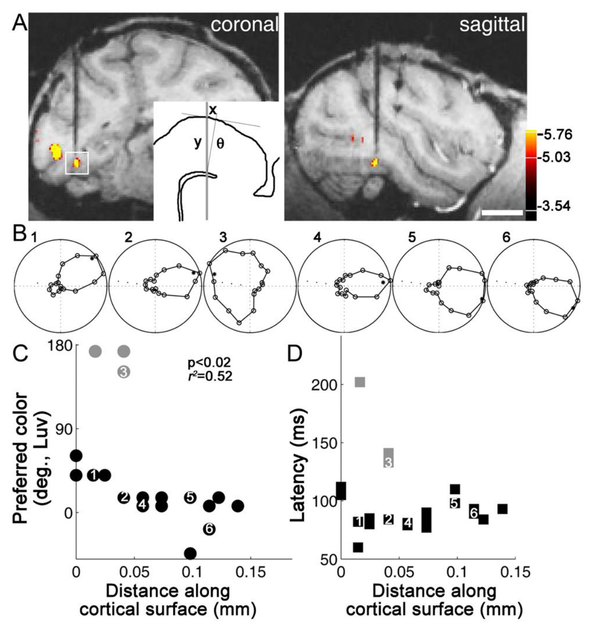
图 10 · PIT 蓝色调细胞的饱和度调谐与潜伏期编码
原文图注:Figure 10. Responses of a blue-tuned cell in PIT assessed using stimuli defined by the cone-opponent axes with which the retina/LGN represent color. A, Cone-opponent "DKL" color space, showing colors in the equiluminant plane at the adaptation point. B, Color stimuli of various saturations and luminance contrasts defined by the cone-opponent color space and projected in the standard C.I.E. xyY chromaticity space. C, Response of one color-tuned neuron to stimuli of varying saturation. Latencies (symbols) are systematically longer with lower saturation stimuli (responses at each saturation averaged across colors; colors defined within the equiluminant plane defined by the adapting background). Inset shows color tuning measured at three saturation levels. Figure prepared by Monica Gates and Galina Gagin (Unpublished data of the author).
(A, B) DKL 与 C.I.E. xyY 色度空间中的刺激定义;(C) 神经元在不同饱和度刺激下的响应潜伏期,表现为较低饱和度刺激对应系统性延长的放电潜伏期,插图显示不同饱和度下色相调谐曲线基本稳定。
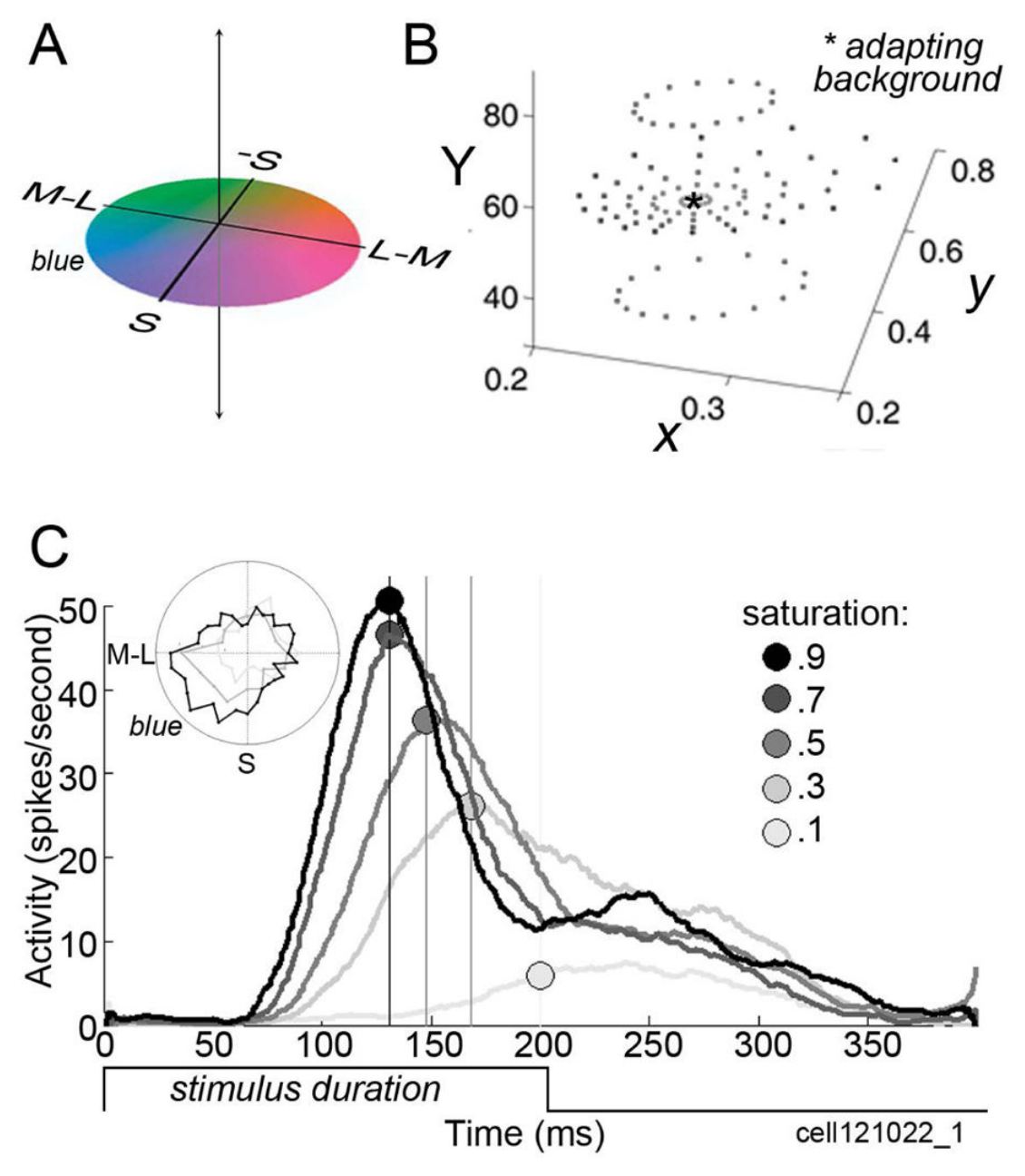
图 11 · Munsell 知觉颜色空间系统
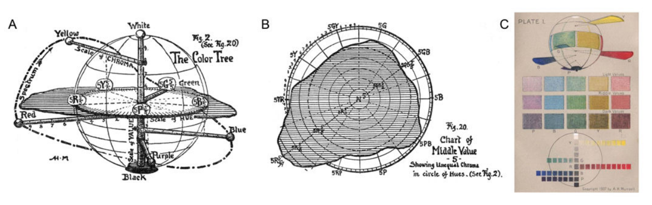
原文图注:Figure 11. Munsell color system of perceptual color space. A, From Munsell (1907): "The color tree is made by taking the vertical axis of the [color] sphere, which carries a scale of value [brightness or luminance], for the trunk. The branches are at right angles to the trunk; and, as in the sphere, they carry the scale of chroma [saturation]. Colored balls on the branches tell their Hue. In order to show the MAXIMA of color, each branch is attached to the trunk (or neutral axis) at a level demanded by its value,—the yellow nearest white at the top, then the green, red, blue, and purple branches, approaching black in the order of their lower values. The color tree prolongs [the chroma axes to represent] the most powerful red, yellow, green, blue, and purple pigments which we now possess, and could be lengthened, should stronger chromas be discovered." B, A horizontal plane through the Munsell system showing the non-spherical nature of the color plane. C, A color plate from Munsell's original book, showing The color sphere (top), fifteen typical steps taken from the sphere (middle), and the value and chroma scale (bottom). As Munsell noted, "Pigment inequalities here become very apparent."
(A) Munsell 颜色树结构(主干为明度,水平分支为彩度/饱和度);(B) Munsell 系统的水平色相平面非对称几何结构;(C) 历史文献中的 Munsell 颜色球及各梯度样本图版。
讨论
作者结合实验证据提出,视觉系统在皮层下阶段利用群体编码表达外周拮抗信号,而在纹外皮层通过区间编码提炼出针对特定色相的调谐单元。在此框架下,S 视锥信号在两条主要视觉通路中发挥不同功能:在腹侧通路后下颞叶的颜色偏好区(globs),S 输入用于实现色调空间的完整均衡表达;在背侧 MT 区及腹侧非颜色偏好区,S 信号提供与亮度解耦的环境光照与阴影变化线索,辅助宏观场景分析与运动感知。
文章指出了当前研究的局限性:V1 日光轴偏置在人类与猕猴 fMRI 结果中存在物种表现差异;LGN 直接投射至 MT 的粒状细胞生理特性有待进一步记录;此外,PIT globs 区域是否构成主观色调知觉的因果中枢,仍需结合更密集的色空间采样与神经干预手段进行验证。
一句话总结
本综述分析了 S 视锥信号在背侧与腹侧通路的功能分化,提出其在腹侧颜色特化区参与知觉色调空间的构建,而在背侧通路及非颜色区主要提供环境光照与阴影线索以支持三维空间几何及运动感知。