该研究探讨下颞叶(inferior temporal, IT)皮层神经元在不同刺激维度上的调谐宽度是否存在内在关联:对特定形状表现出稀疏放电(高选择性)的神经元,在尺寸、位置、朝向及纹理等维度上是否同样表现出较窄的调谐带宽。作者记录了两只猕猴前部 IT 皮层单单元对基准剪影、连续平滑变形系列及恒等保持几何变换的电生理响应,发现神经元的稀疏度指标在各测试维度间均呈显著正相关,表明调谐锐度具有跨维度的内在一致性。
研究背景
IT 皮层神经元对复杂形状的选择性具有高度多样性。既往研究形成了两种调谐特征的经验观察:一是采用多样化自然图像测试时,部分神经元表现为稀疏放电(仅对少数几个刺激产生响应),部分神经元则表现为广泛分布的梯度响应(Gochin 等,1994;Tamura & Tanaka,2001);二是在连续参数化刺激空间中,单细胞调谐曲线的宽度存在从窄到宽的连续分布(Op de Beeck 等,2001;Zoccolan 等,2007)。
关于这两类表征特征的组织模式存在两种假设:异质性假说认为调谐带宽取决于刺激特征与感受野局域特征的随机重叠程度,预测不同参数维度之间的调谐宽度缺乏系统相关;内在特性假说则认为神经元存在统一的调谐锐度倾向,调谐宽度在不同刺激属性间呈跨维度正相关。在初级视皮层(V1),神经元的朝向选择性带宽与空间频率带宽已有正相关报道;而在 IT 皮层,高选择性神经元往往对尺寸和位置变化表现出更低的放电耐受度(Zoccolan 等,2007)。要厘清调谐宽度是由输入特征偶然决定还是由神经元内在特性约束,需要在同一批细胞中系统比较离散形状稀疏度与多维参数调谐宽度的对应关系。
研究思路
实验以 8 个标准化剪影图形作为基准刺激集,沿两个正交方向扩展测试维度:其一,将基准形状两两配对,沿非均匀有理 B 样条(NURBS)控制点生成连续变形序列(morph lines),检验形状拓扑渐变下的调谐形态;其二,对每个基准形状施加不同水平的尺寸、空间位置与平面旋转等恒等保持变换。
分析采用稀疏度(sparseness)指标(取值介于 0 至 1 之间,0 代表对所有刺激均等放电,1 代表仅对单一刺激响应)量化各维度调谐锐度。若异质性假说成立,不同变形序列或几何变换下的稀疏度应彼此独立;若内在特性假说成立,神经元在各维度上的稀疏度应呈显著正相关。作者进一步通过基线漂移校正、电生理波形特征排查、解剖记录位置检验,以及追加的自然纹理刺激集测试,系统评估这种跨维度关联的普遍性。
方法
两只成年雄性帽猴(编号 Ka 与 Sa,约 7 岁)参与实验。结构 MRI 确认记录位点位于左侧前部 IT 皮层。被试在红外眼动监测下完成中央注视任务(注视窗口 3°,平均注视误差标准差小于 0.33°,注视达标后给予果汁奖励)。采用 24 通道线性微电极阵列记录细胞外放电,经离线聚类严格筛选出孤立的单单元信号。最终纳入 99 个对变形序列及几何变换均具备稳定视觉响应的单神经元(Ka 猴 54 个,Sa 猴 45 个)。
主实验共呈现 116 个刺激条件,每个重复 8 次:包括 8 个基准白色剪影(视角 3°);4 条各包含 11 个刺激的连续变形序列(共 44 个图形);以及基准图形在尺寸(1.5°、3°、4.5°、6°)、外周偏心位置(0°–4.5°)与平面朝向(0°–90°)各 3 个变化梯度的几何变换组。神经放电率取刺激呈现后 50–300 ms 窗内均值。子集分析对 Ka 猴的 28 个神经元额外记录了 80 个独立剪影与 480 种自然纹理图案。容忍度分析区分了两类指标:绝对容忍度(反应幅值随变换波动的稳定性)与相对容忍度(形状间相对偏好顺序跨变换维度的向量相关度)。
主要结果
- 形状变形序列间的调谐锐度高度相关:99 个神经元在不同变形序列间的稀疏度呈显著正相关(如序列 3 与序列 4 稀疏度相关系数 r=0.70,P<0.000005;图 3A);基准集稀疏度与最大变形序列稀疏度相关系数达 r=0.90(P<0.000005;图 3B),基准集与各单条序列间的两两相关均达到统计学显著水平(图 3C)。扣除自发基线放电后上述相关依然稳固(序列间平均 r=0.56)。
- 单细胞调谐的一致性表现:稀疏放电的典型神经元在各条变形序列以及尺寸、位置变化下均呈现较窄的调谐带宽,而分布放电的神经元在各个测试序列中均维持平缓宽泛的调谐轮廓(图 2、图 5)。
- 扩展至恒等保持几何变换:尺寸变换稀疏度与基准形状稀疏度显著相关(r=0.76,P<0.000005;图 6),空间位置与旋转朝向的稀疏度同样与基准集呈现显著正相关。
- 绝对容忍度与相对容忍度的分离表征:采用绝对容忍度定义时,神经元稀疏度与尺寸/位置容忍度呈强负相关(r=−0.81 与 −0.77,P<0.000005),复现了既往关于高选择性细胞放电幅值波动较大的报道;但当采用相对容忍度(形状偏好排序的保持程度)时,稀疏度与容忍度呈正相关(尺寸变换 r=0.55、位置变换 r=0.39、朝向变换 r=0.33,P<0.005;图 7),表明高选择性神经元在几何变换下能更稳定地保持其固有的形状偏好关系。
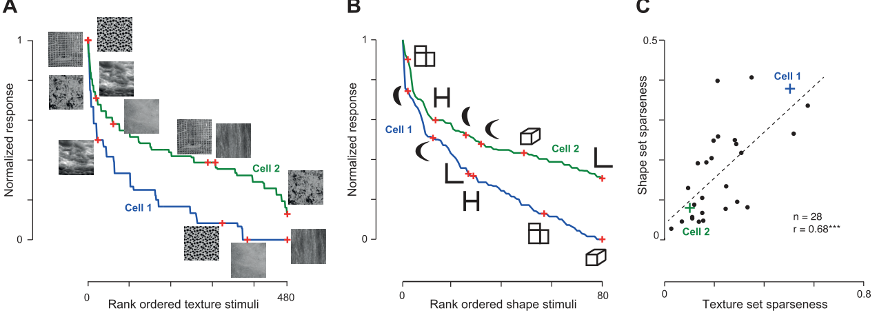
- 形状与自然纹理维度的通用性:在 28 个同时接受两类测试的神经元中,独立剪影集与 480 张自然纹理集的稀疏度呈显著正相关(r=0.68,P<0.0005;图 8)。神经元调谐稀疏度与动作电位波形宽度、最大放电率及前后/内外解剖记录坐标均未检测到显著相关性(|r|≤0.14)。
图注解读
图 1 · 两种选择性图景的逻辑对照
原文图注:Fig. 1. Possible relationships between selectivity across shape space. Consider 2 inferior temporal (IT) neurons: the first neuron is a sparse firing neuron that responds to 2 distinct stimuli (left). The second neuron is a distributed firing neuron that responds to 4 distinct stimuli (middle). Each row represents a scenario that shows how each neuron might respond to small variations in the neighborhood of its preferred stimuli. Top row: Scenario 1: selectivity is heterogeneous. Neuron 1 is sharply tuned to changes in stimulus 1 and broadly tuned to variations around stimulus 2, whereas neuron 2 is sharply tuned around both stimuli. …
该图示意两种假设模型:上行展示异质性假说,即同一神经元在其偏好刺激 1 周围表现为窄调谐,而在刺激 2 周围表现为宽调谐,不同维度间缺乏固定对应关系;下行展示内在特性假说,即神经元在各个偏好刺激周围均表现出一贯的窄调谐或一贯的宽调谐,提示调谐宽度在各刺激维度间具有协变关联。
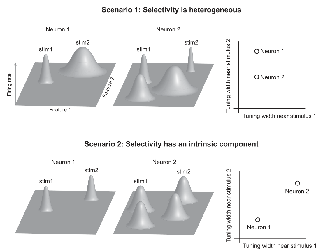
图 2 · 一稀一宽两个示例神经元
原文图注:Fig. 2. Example responses to the reference set (A) and to morphed variations (B–E). The first neuron (red rasters) fires sparsely to shapes in the reference set and has sharp tuning to smooth parametric variations of each stimulus. The second neuron (blue rasters) shows graded firing to all stimuli in the reference set and also shows broad tuning to smooth parametric variations. Each box depicts the individual spikes elicited by that stimulus and the histogram with the average firing rate across trials in bins of 10 ms each.
该图展示 2 个典型神经元的放电栅格图与反应直方图:红色标记细胞在基准集(A)中仅对少数剪影放电,且在 4 条连续变形序列(B–E)中呈现陡峭的调谐曲线;蓝色标记细胞对基准集呈连续阶梯放电,并在所有变形序列中维持平缓宽阔的调谐曲线,直观呈现了单细胞水平的调谐宽度差异。
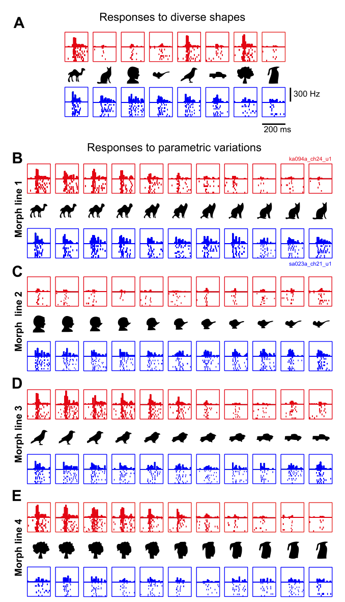
图 3 · 变形线之间的稀疏度正相关
原文图注:Fig. 3. Highly selective neurons are selective along many stimulus variations. A: sparseness along morph line 4 plotted against sparseness along morph line 3 for each neuron. Action potential waveforms (black lines within green, red, and blue shaded areas indicate means; green shaded areas indicate SD) are shown for a subset of neurons (green dots) to avoid crowding. The red and blue squares correspond to the red and blue example neurons shown in Fig. 2. B: sparseness on the reference set (excluding the morph pair that gave maximum sparseness) plotted against maximum morph line sparseness across neurons with conventions as before. C: matrix of pairwise correlations between reference set sparseness and sparseness along each individual morph line. All correlations were statistically significant (*P < 0.000005).
子图 A 展示神经元在序列 3 与序列 4 稀疏度散点图(r=0.70),附带典型动作电位波形;子图 B 展示基准形状稀疏度对最大变形序列稀疏度的相关图(r=0.90);子图 C 展示基准集与各单条序列稀疏度的两两相关系数矩阵,所有相关均达显著水平(P<0.000005),支持跨变形维度的调谐宽度一致性。
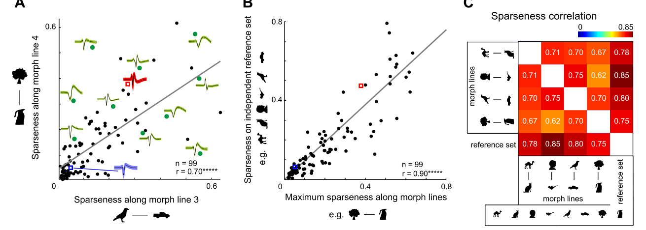
图 4 · 选择性与波形、放电率和记录位置无关
原文图注:Fig. 4. Relationship between sparseness and other intrinsic properties. A: overall sparseness for each neuron (on the entire stimulus set) plotted against the width of its action potential (peak-to-trough time). Red and blue dots correspond to neurons from monkey Ka and monkey Sa, respectively. The correlation coefficient is indicated in the plot (n.s., not statistically significant). B: overall sparseness for each neuron plotted against its maximum firing rate (across all stimuli). C: overall sparseness for each neuron plotted against its recording location along the anterior-posterior (AP) axis relative to the center of the recording chamber for each monkey. D: overall sparseness for each neuron plotted against its recording location along the medial-lateral (ML) axis relative to the center of the recording chamber for each monkey.
该图检验潜在电生理与解剖混杂变量:四个散点图分别展示总体稀疏度与动作电位峰谷宽度(A)、最大放电率(B)、前后轴记录坐标(C)及内外轴记录坐标(D)的散点分布。各项相关均无统计学显著性,排除了细胞类型或解剖梯度偏置对调谐宽度的系统性影响。
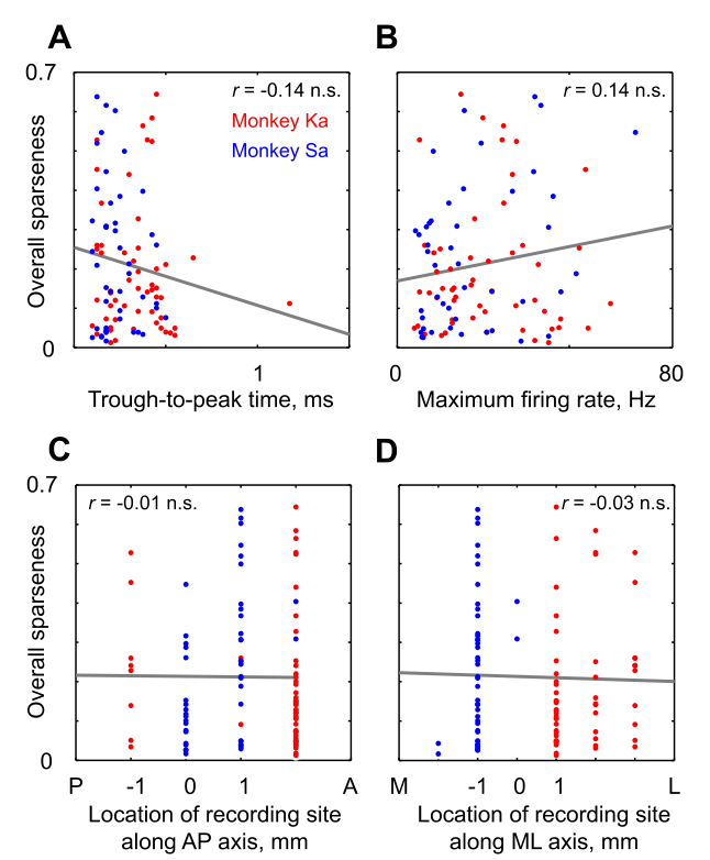
图 5 · 尺寸、位置、朝向下的示例反应
原文图注:Fig. 5. Example responses for size, position, and orientation variations. The neurons are the same as depicted in Fig. 2. To examine tolerance to identity-preserving transformations, each shape in the reference set was presented at 3 additional sizes, positions, and orientations. The sparse neuron (red rasters) shows greater modulation to changes in size, position, and orientation, whereas the distributed neuron (blue rasters) shows gradual changes. Conventions are as before.
该图展示图 2 中两个示例神经元在尺寸、偏心位置及朝向变换下的放电表现。稀疏放电细胞(红色)在各类变换下放电幅值调制明显,而宽调谐细胞(蓝色)随变换参数呈现较为平缓的变化,为后续计算两类容忍度指标提供了实验数据基础。
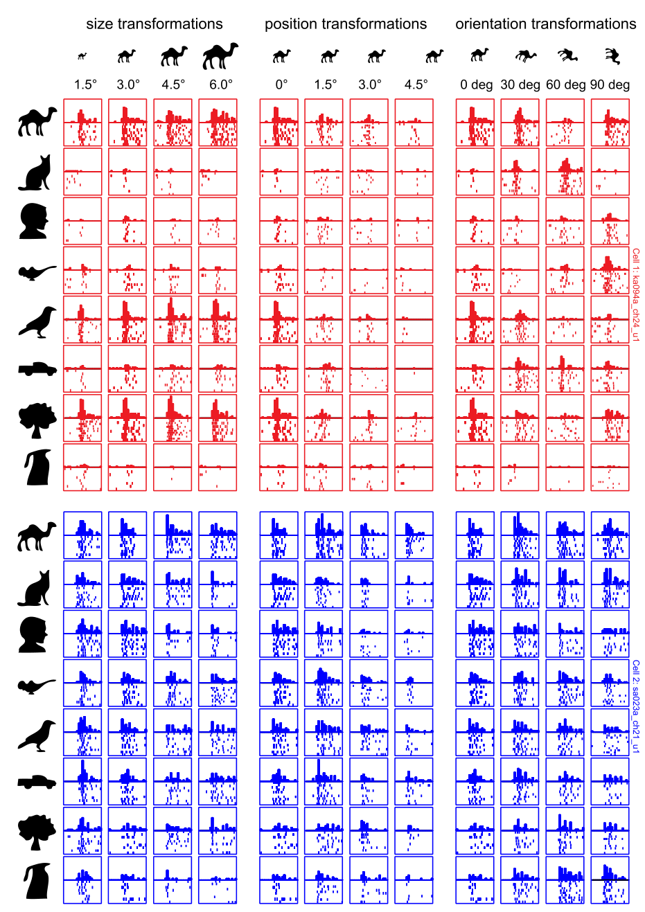
图 6 · 尺寸/位置/朝向选择性也相关
原文图注:Fig. 6. Highly selective neurons are also selective for size, position, and orientation. A: sparseness along size variations plotted against sparseness on the reference set for each neuron. B: matrix of pairwise correlations between sparseness on the reference set with average sparseness along size, position, and orientation variations. All correlations were statistically significant (*P < 0.000005).
子图 A 展示尺寸变换稀疏度对基准集稀疏度的相关散点分布(r=0.76);子图 B 展示基准集与尺寸、空间位置、朝向各变换稀疏度的两两相关系数矩阵(均显著,P<0.000005)。表明对形状特征高度选择的神经元,对视网膜投射变换同样表现出较为敏感的调谐特性。
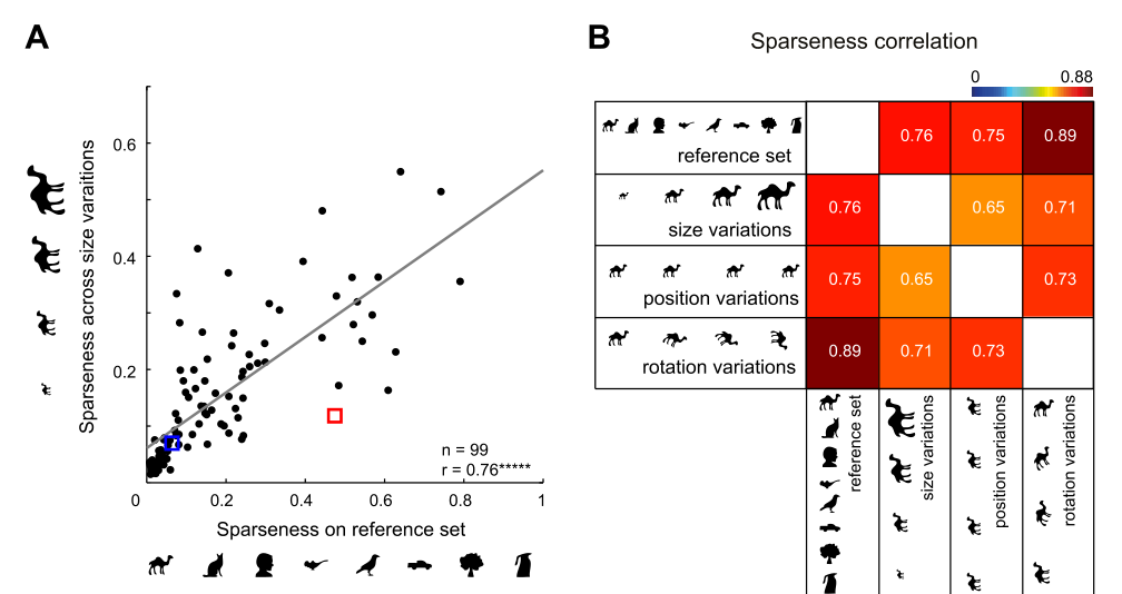
图 7 · 绝对容忍度负相关、相对容忍度正相关
原文图注:Fig. 7. Relationship between tolerance and selectivity in IT neurons. A: absolute size tolerance for each neuron plotted against reference set sparseness. The absolute size tolerance is a measure of how much the neural response changes with changes in stimulus size (see MATERIALS AND METHODS). B: relative size tolerance for each neuron plotted against reference set sparseness. Relative tolerance is a measure of how strongly a neuron maintains its shape preferences across changes in size (see MATERIALS AND METHODS). C: summary of correlations of reference set sparseness with relative tolerance and absolute tolerance. P < 0.005; P < 0.0005; ****P < 0.000005.
子图 A 呈现绝对尺寸容忍度与基准稀疏度的负相关(r=−0.81);子图 B 呈现相对尺寸容忍度与基准稀疏度的正相关(r=0.55);子图 C 汇总了三种几何变换下两类容忍度与稀疏度的相关系数对比。结果表明高稀疏度细胞放电幅值易受几何变换调节,但其对形状刺激的相对偏好构型在变换下能够较好维持。
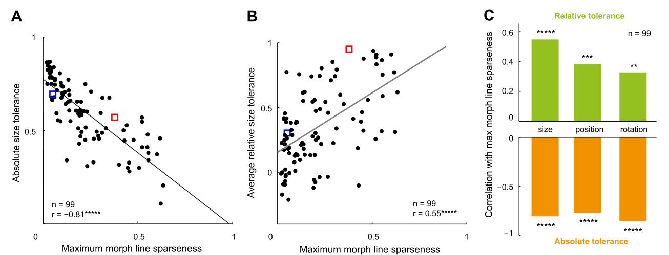
图 8 · 形状选择性与质地选择性相关
原文图注:Fig. 8. Selectivity for shape and texture across neurons. A: example responses for 2 IT neurons to a set of natural textures. The normalized responses of each cell are rank ordered from best to worst, with sample stimuli shown along the tuning curve. Blue curve, first neuron; green curve, second neuron. B: example responses for the same 2 neurons to a set of silhouette shapes. C: sparseness for the shape set plotted against the sparseness of the texture set across the sampled neural population (n = 28). The example neurons are highlighted using crosses. ***P < 0.0005.
子图 A 与 B 分别展示 2 个典型神经元对 480 张自然纹理与 80 张剪影图形的排序反应曲线;子图 C 展示 28 个神经元在形状集稀疏度与纹理集稀疏度之间的相关散点图(r=0.68,P<0.0005),证实跨越不同刺激属性维度的调谐宽度一致性。
讨论
研究结果支持 IT 皮层神经元存在全局性的调谐锐度约束。单细胞并非随机地在某些维度呈现窄调谐而在其他维度呈现宽调谐,而是展现出系统性的跨维度协变,这一规律在形状连续变形、仿射几何变换以及非形状纹理维度上均得到验证。此类跨维度调谐特征与 V1 皮层关于朝向和空间频率带宽的相关性研究具有类比性。
关于选择性与容忍度(不变性)的关系,分析表明区分绝对容忍度与相对容忍度具有方法学意义:高稀疏度神经元在绝对放电幅值上受几何参数变化调节较为明显,但在相对形状偏好排序上表现出更高的保真度,表明锐调谐神经元能够在几何变换干扰下稳定编码物体身份。作者探讨了内在锐度的潜在生理学基础,排除了动作电位波宽及大尺度解剖梯度的影响,推测局域抑制性中间神经元回路或特异性前馈汇聚可能共同决定了单细胞的调谐锐度。研究局限包括记录范围主要集中在左前部 IT 皮层,所用刺激主要为二维轮廓剪影与静态纹理,未来有待在三维立体线索及主动行为任务中进一步验证。
一句话总结
该单细胞电生理研究表明,猕猴下颞叶神经元的调谐带宽在连续形状变形、尺寸位置变换及自然纹理等多维度上表现出显著正相关,且高选择性神经元能够更好地在几何变换中维持相对形状偏好,表明单细胞调谐锐度受内在共性约束。