一句话定位:这项解剖示踪研究在 7 只恒河猴中结合微电极电生理测绘与多种顺向、逆向示踪技术,系统解析了后部下颞叶皮层(TEO)在不同视野离心度上的皮层连接网络与层板组织模式,确立了 TEO 作为枕颞腹侧通路中继站的解剖学地位,并揭示了中央凹与周边视野代表区在投射网络上的差异。
研究背景
下颞后部皮层 TEO 是理解腹侧视觉通路的关键节点。细胞构筑学早已把它与前方 TE 区分开,行为损毁又提示两者功能不完全相同:TEO 切除主要影响二维图形辨别,TE 切除则更严重地影响物体再认记忆。随后电生理测绘发现,TEO 含有对侧视野的完整但粗糙的视网膜拓扑代表,中央视野位于外侧凸面,周边视野延伸到腹侧面和枕颞沟附近。这一结果说明 TEO 不是一块均质皮层,要理解它的输入输出,注射位置必须对应实际视野代表区,不能只按脑表面的大致边界操作。
此前的连接研究却主要集中在 TEO 背侧,也就是中央视野代表区,而且很少同时检查顺向传出与逆向传入。因而仍不知道它与 V2、V3、V4、TE、上颞沟、顶叶和内侧颞叶的完整连接格局,也不清楚各连接在皮层层次上的起源和终止。层板分布很重要,因为前馈投射通常由粒上层发出并重点终止于第 IV 层,反馈投射则呈不同层型;只有区域清单而没有层型,就很难判断 TEO 在加工层级中的方向。中央与周边代表区是否接入不同脑区也未被检验,这关系到高分辨中央视觉与外周空间信息是否沿不同路线处理。
本文因此把生理定位、小体积注射和双向示踪结合起来,系统补齐这张连接图。一个具体动机是解释旁路:若 V4 损毁后 TE 仍有视觉反应,TEO 或其他区域可能提供绕过 V4 的输入,但必须先知道 TEO 实际与哪些区域相连。研究还借意外注入 PITd 的例案比较连接差异,以检验生理边界是否有解剖对应。所得结果用于建立腹侧通路的底图,而不是单凭连接就证明某条信息流负责识别;它提供的是后续功能与损毁研究必须解释的解剖约束。
研究思路
研究采用“微量注射、全视野覆盖、双向示踪、层板解析”的技术路线。通过术中电生理测绘准确标定 TEO、V4 及 TE 的功能边界与局部视野表征偏心度,将微量顺向与逆向示踪剂分别注入 TEO 的中央凹代表区、上视野周边代表区与下视野周边代表区,以对比不同离心度的投射模式。
在连接方向的判别上,依据皮层投射的层板终止规则(laminar rules):前馈连接表现为起源于粒上层(II/III 层)且轴突终末密集分布于靶区第 IV 层;反馈连接表现为起源于粒下层(V/VI 层)且终末主要避开第 IV 层;中间型(intermediate-type)连接表现为起源细胞在粒上与粒下层均匀分布,且终末在皮层各层均有分布。为排除过路纤维吸收伪影,仅将在至少 40% 的测试个案中稳定复现的投射判定为可靠连接;并利用一例误注入后下颞背侧区(PITd)的对照个案,检验 TEO 与相邻脑区的解剖特异性。
方法
实验使用 7 只雄性恒河猴(体重 3–6 kg)。每只猴在无菌手术下植入记录舱,在氯胺酮镇静与人工通气麻醉下进行两次微电极记录(间隔 5 天),通过多单位放电测绘确定 TEO 及毗邻脑区的视网膜拓扑分布,并确定注射深度与坐标。微量注射利用 1 μl Hamilton 注射针以 0.01–0.1 μl/min 的流速执行。逆向示踪剂采用快蓝(Fast Blue, FB)、双脒黄(Diamidino Yellow, DY)、罗丹明标记葡聚糖(Rhodamine Dextran, RD)及辣根过氧化物酶结合麦胚凝集素(WGA-HRP);顺向示踪剂采用 WGA 及氚标记氨基酸混合物([3H]脯氨酸与[3H]亮氨酸等比例混合)。注射体积控制在 0.1–0.2 μl 之间。
动物灌注后取脑制作 50 μm 连续冷冻切片(冠状或矢状切片,间隔 250 μm),交替进行荧光显微镜检、放射自显影处理(曝光 20 周)、WGA-HRP 显色反应、Gallyas 髓鞘染色及硫堇尼氏染色。利用计算机重建系统沿第 IV 层轮廓重构大脑二维皮层展开图(unfolded cortical map),定量统计 26 个候选视皮层区的标记分布。共获得 11 处 TEO 注射位点:2 处位于中央凹代表区(偏心度 <1°)、5 处位于上视野周边区(偏心度 10°–22°)、2 处位于下视野周边区(偏心度 17° 与 26°)、1 处位于水平子午线区(偏心度 4°)、1 处位于 23° 下周边区;另包含 1 处误注入 PITd 的个案,形成 15 个 TEO 实验例案(7 顺向,8 逆向)和 1 个对照例案。
主要结果
-
TEO 的主要前馈输入源于 V2、V3 及 V4 并呈现明确的拓扑组织:注射位点周围标记的细胞主要分布在相应视网膜拓扑区域(如中央凹注射标记 V2 与 V4 的中央代表区,上/下视野注射分别标记腹侧或背侧的 V2/V3/V4 代表区),连接范围延伸至至少 25° 偏心度(图 2、图 4、图 8、图 11)。所有输入区域均接受来自 TEO 的反馈投射;其中与 V4 的双向连接密度最高。全样本中均未观察到 TEO 与初级视皮层(V1)之间的直接联系。
-
较稀疏的附加前馈输入来自背侧/运动相关区:包括 V3A、V4t 与 MT 区域;该类连接密度较低,且主要出现在注入 TEO 周边视野代表区的情形中——V3A 仅与偏心度大于 4° 的区域相连,V4t 与顶叶 PP 区亦仅在周边注射后被标记(图 7、图 8、图 12)。
-
TEO 与顶叶及额叶眼区形成中间型双向连接:TEO 与 FST、顶下小叶内侧的 PP 区、顶内沟外侧顶区(LIP)、额叶眼区(FEF)以及海马旁回 TF 区存在相互的中间型连接;标记神经元呈双层分布,轴突终末分散于各层但未在第 IV 层形成聚集(图 8、图 10、图 14)。TEO 与 LIP 之间的连接未表现出视网膜拓扑组织。
-
前馈输出主要投向前颞下回(TE)与上颞沟前部:TEO 产生密集的第 IV 层轴突终末前馈投射,主要投向 TE、TEm/TEa 及 IPa,并接收来自这些区域的反馈回路(图 5、图 6D、图 12)。此外,反馈投射还发自海马旁回 TH、颞极 TG 及鼻周皮层 36 区。中央凹注射未观察到向 TF/TH 的明显输出,而周边视野注射则形成投射。
-
中央凹与周边视野投射差异及与 PITd 的界限:中央凹注射完全未标记 V3A、V4t、PP 与 7a 区,而周边视野注射系统性标出上述脑区以及顶内沟后段区域。误注入相邻 PITd 的对照例案展现出与 TEO 相似的整体投射,但 V2 与 V3 区域完全无标记(图 13),证实了 TEO 与 PITd 在解剖连接模式上的独立性。对侧半球对称位点主要通过粒上层细胞介导连合投射。
图注解读
图 1 · 皮层展开图上的视觉区分布与 TEO 拓扑
原文图注:Fig. 1. Location of visual areas identified in occipital, parietal, and temporal lobes of macaque monkeys. Areas are shown on a two-dimensional unfolded cortical map at the right (C; adapted from Ungerleider and Desimone, '86b). Striate cortex is not included. Thick lines represent the boundaries of sulci; thin lines at the perimeter of the map indicate where the map was "cut" from the rest of the cortex. ……
展示恒河猴枕叶、顶叶和颞叶视皮层的解剖分区方案:左侧为侧视图,右侧 C 为剔除 V1 后的二维皮层展开图(粗线表示脑沟底界,细线表示外切边缘),D 展示 TEO 的视网膜拓扑组织(星号标出中央凹,加减号标出上下视野,VM/HM 标示垂直与水平子午线)。为后续示踪分析提供了统一定位模板。
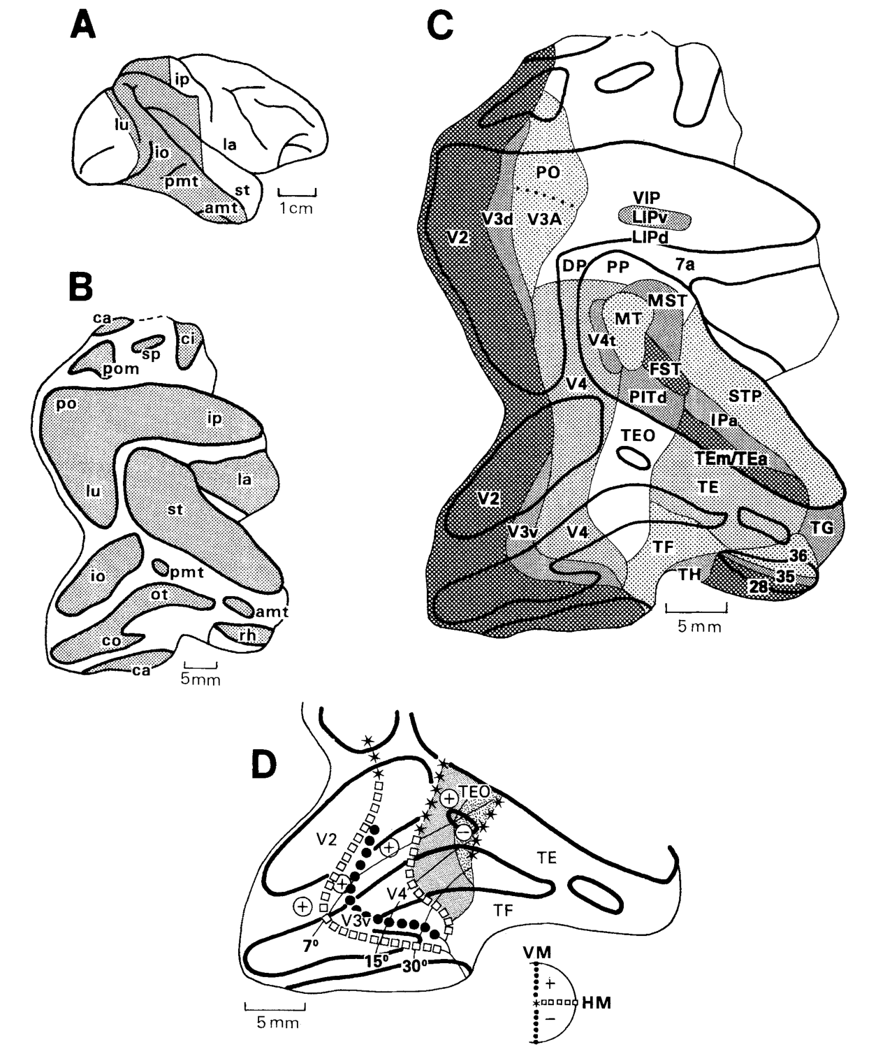
图 2 · 中央凹 TEO 注射后的逆向标记(侧视/底视/冠状切片)
原文图注:Fig. 2. Case 1-WGAr: Distribution of retrogradely labeled cells after wheat germ agglutinin (WGA) injection into the foveal representation of TEO. The receptive field recorded at the injection site is shown at the right. Injection site (black), diffusion halo (hatched), and labeled cells (dots) are shown on the lateral (upper left) and ventral views (lower right), as well as on representative coronal sections (#1-5). ……
中央凹代表区(偏心度 <1°)注入 WGA 后的逆向标记分布图,右侧给出注射位点的感受野大小与位置。黑点标示标记细胞分布:显著标记出现在 V2/V3/V4 的中央凹代表区、STS 尾部的 MT 与 FST、PITd、STS 前部的 TEm/TEa 与 IPa,以及 TE、TF、TH、TG、36 区与 LIP;V4t 与 PP 等区域无标记,为主要结果 1 和 4 提供支持。
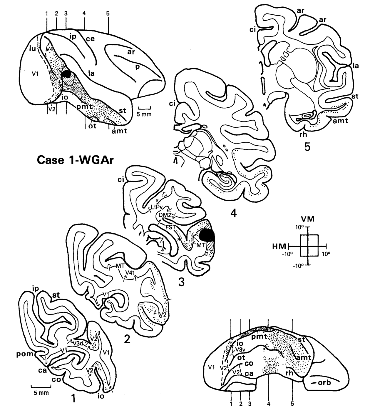
图 3 · 同一例案在展开图上的标记分布
原文图注:Fig. 3. Case 1-WGAr: Distribution of retrogradely labeled cells after WGA injection into the foveal representation of TEO, shown on an unfolded map of the posterior part of the cortex. Heavy dashed lines on the map indicate myeloarchitectonic borders. Thin lines are the layer IV contour lines of the same sections shown in Figure 2. The sulcal pattern in this case was unusual in that io and pmt merged to form a single sulcus. ……
将图 2 标记位点重叠于二维皮层展开图上。粗虚线为髓鞘构筑确立的解剖边界,细线为各冠状切片第 IV 层的轮廓投影。标记神经元紧密聚集在 V2/V3/V4 对应的中央凹视野区,支持 TEO 输入投射具有严格的拓扑映射特性。
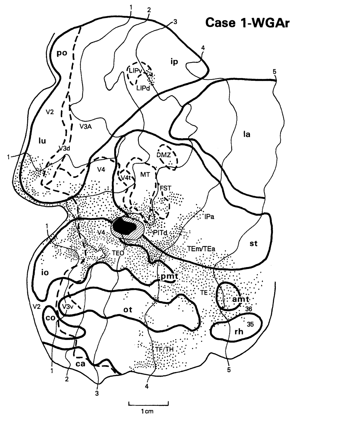
图 4 · 另一中央凹例案(RD 示踪)的重复验证
原文图注:Fig. 4. Case 1-RD: Distribution of retrogradely labeled cells after a dextrane (RD) injection into the foveal representation of TEO, shown on the lateral and ventral views (left) and an unfolded cortical map (right). All conventions are as in Figures 1-3.
在另一更靠前的 TEO 中央凹位点注入罗丹明葡聚糖(RD)的逆向标记验证:V2 与 V3 区域依然出现稳定标记,且未检测到 V1 的直接标记,排除了示踪剂沿针道扩散入 V4 造成的假象,确认了 V2/V3 直投 TEO 通路的存在。
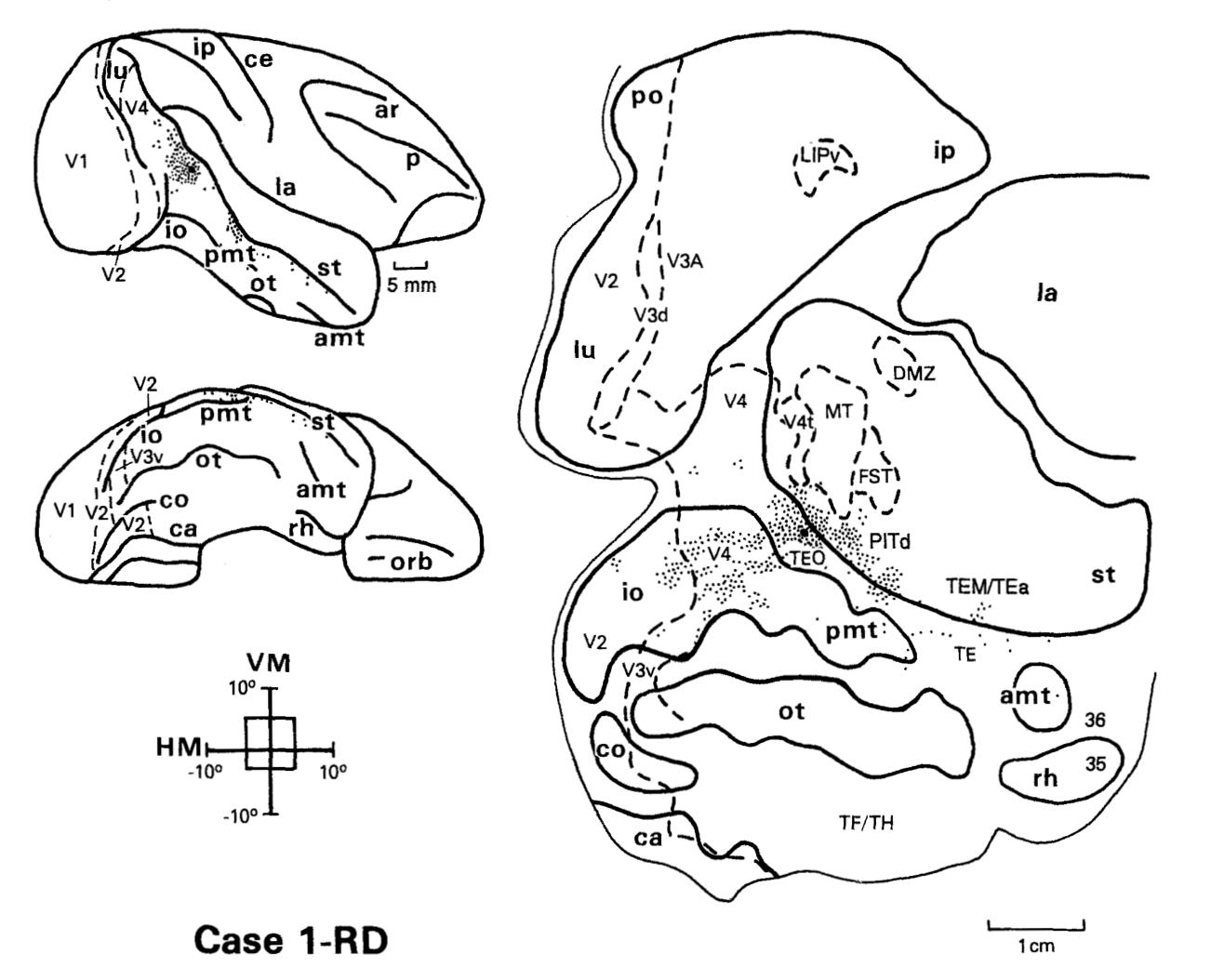
图 5 · 中央凹注射后的顺向终末标记
原文图注:Fig. 5. Case 1-WGAa: Location of anterogradely labeled terminals after the same foveal WGA injection shown in Figures 2 and 3. Projections avoiding layer IV are represented by light shading, while projections occupying all layers or heavier in layer IV are represented by dark shading. All other conventions are as in Figures 1-3.
中央凹 WGA 注射对应的顺向轴突终末层板分布图:浅色阴影代表终末主要避开第 IV 层的反馈型投射(出现在 V2、V3、V4),深色阴影代表主要聚集于第 IV 层的前馈型投射(见于 TE 与 TG 区),全层分布见于 TEm/TEa 与 FST,为确定 TEO 在层级系统中的前馈/反馈方向提供解剖证据。
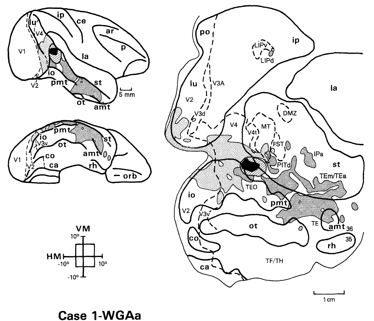
图 6 · 暗视野照片:层型判读的实拍证据
原文图注:Fig. 6. Darkfield photomicrographs show the pattern of retrograde and anterograde label after foveal TEO injection with WGA (case 1-WGA). A, B: Labeling in V2. A is taken from the focus of the labeled region, whereas B is taken from a less densely labeled region. C: Labeling in V4. D: Labeling in TE. The cortical layers (I-VI) and white matter (WM) are indicated. In V2-V4, retrogradely labeled cells occur mainly in the supragranular layers and labeled terminals are lightest in layer IV. In TE, retrogradely labeled cells occur mainly in the infragranular layers and labeled terminals are heaviest in layer IV. Scale bars = 250 μm. ……
暗视野显微照片展示不同靶区的微观标记层板分布:在 V2(A、B)与 V4(C)中,逆向标记细胞主要位于粒上层,顺向终末在第 IV 层明显减弱;在 TE(D)中,标记细胞主要位于粒下层,终末轴突紧密聚集于第 IV 层,确立了前馈与反馈连接判别的显微组织学标准。
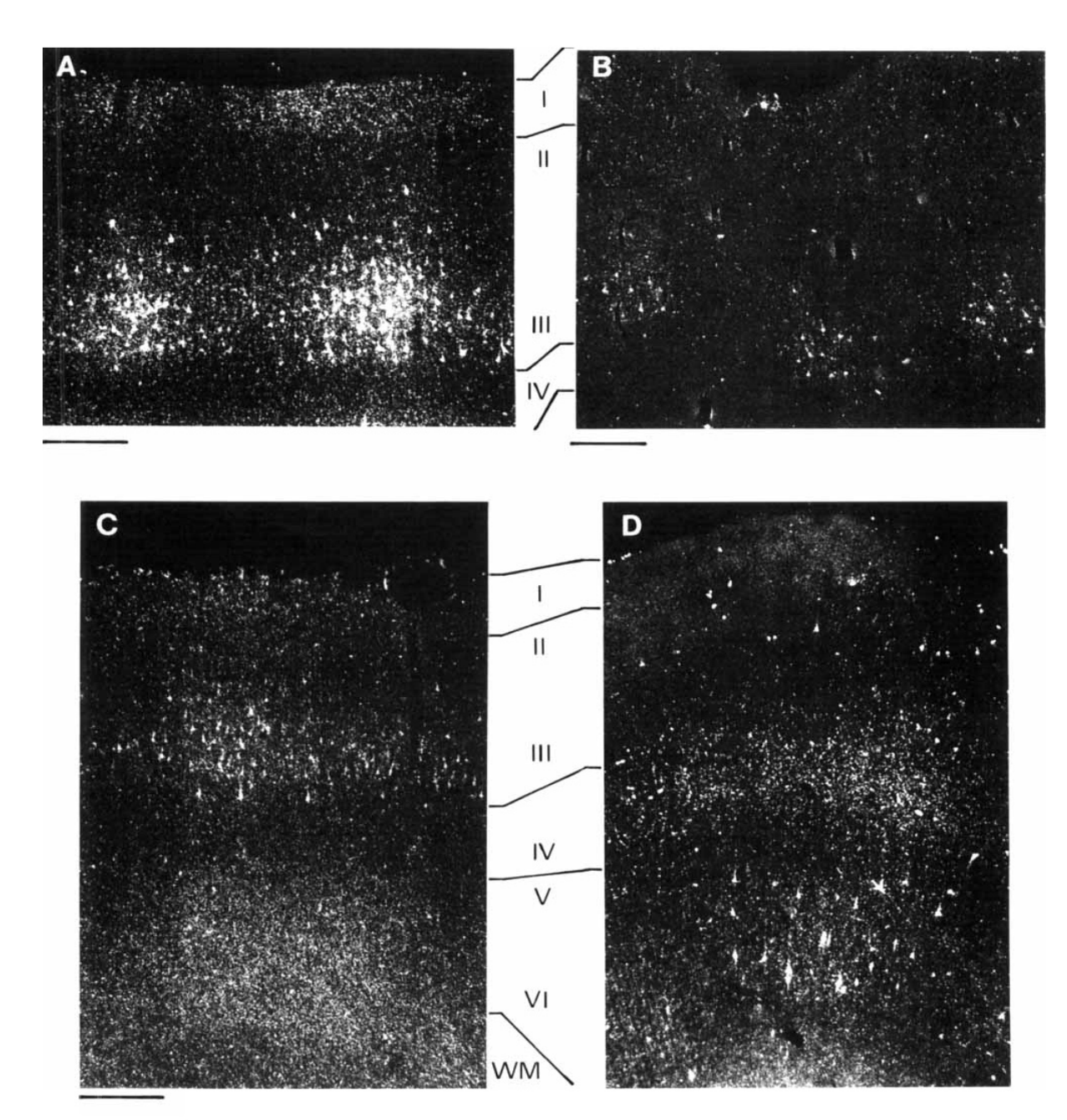
图 7 · 周边视野注射(上下野兼有)的逆向标记
原文图注:Fig. 7. Case 2-WGAr: Location of retrogradely labeled cells after a WGA injection into the periphery of TEO, including both lower and upper visual fields (see text). Arrow on the lateral view of the brain (upper left) points to the location of the injection site buried in the sulcus. Note that MT and V4t appear closer to the lip of st than one might expect (see also Fig. 13). ……
TEO 沟内周边视野代表区注入 WGA 后的逆向标记分布:除常规视区外,新增了 V3A、DP、V4t、PP 以及顶内沟后段的标记细胞,直观展示了周边视野投射网络相比中央凹代表区的广泛性。
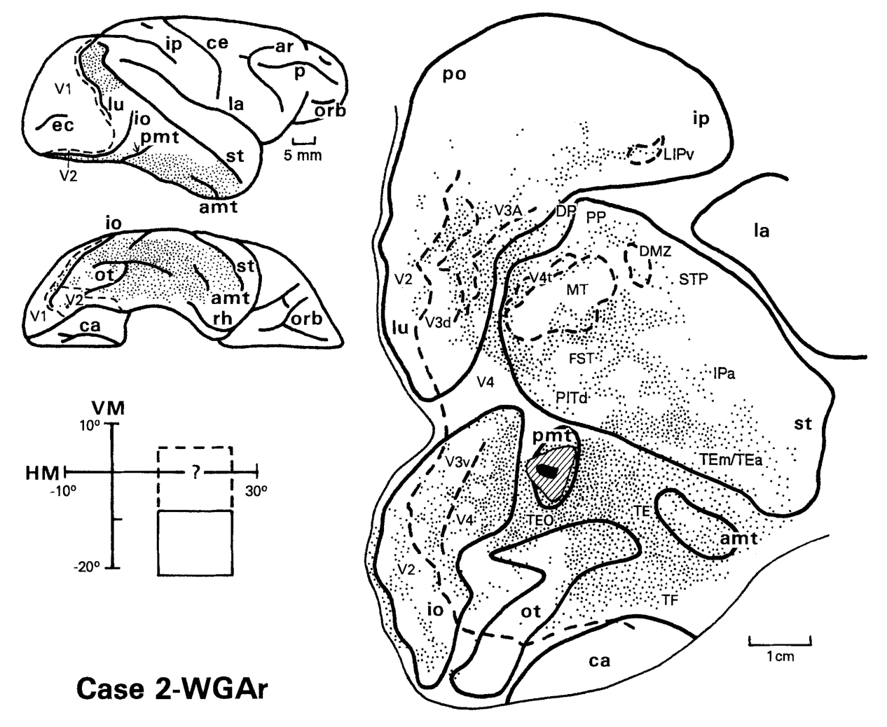
图 8 · 上视野代表区注射(DY)的逆向标记
原文图注:Fig. 8. Case 3-DY: Distribution of retrogradely labeled cells after a diamidino yellow (DY) injection centered in the upper peripheral field representation of TEO. Data are shown on the lateral (upper left) and ventral (lower right) views of the injected hemisphere as well as on representative parasagittal sections (#1-3). Arrows on parasagittal sections mark myeloarchitectonic boundaries of visual areas. Arrow on the ventral view of the brain points to the location of the injection site buried in the sulcus. All conventions are as in Figure 2.
上视野周边代表区(偏心度 22°)注入双脒黄(DY)后的逆向标记分布:标记细胞对应分布于 V2、V3、V4 的腹侧(上视野)皮层,MT 内标记集中于其后内侧(上视野区),并伴随顶叶 7a 区的显著标记,验证了上视野投射的拓扑对应关系。
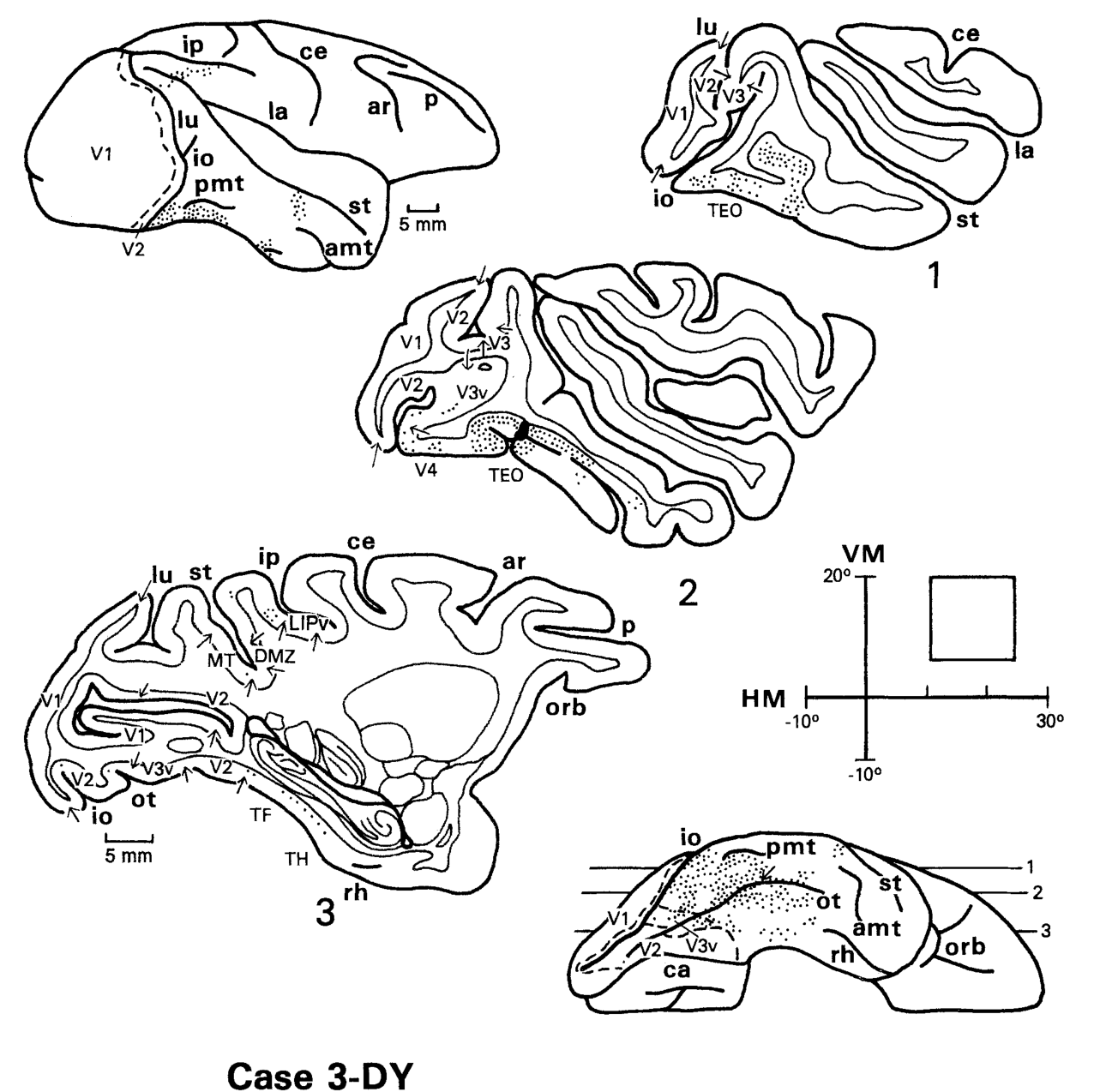
图 9 · 上视野例案的展开图版本
原文图注:Fig. 9. Case 3-DY: Distribution of retrogradely labeled cells after a DY injection centered in the peripheral upper field representation of TEO, shown on an unfolded map of the posterior part of the cortex. Conventions are as in Figure 3.
将图 8 上视野注射的标记映射于皮层展开图,标记区域系统性对应于腹侧早期视区带及 MT 内部的上视野表征带,中央凹区域保持空白,直观展现视野偏心度在皮层连接中的空间一致性。
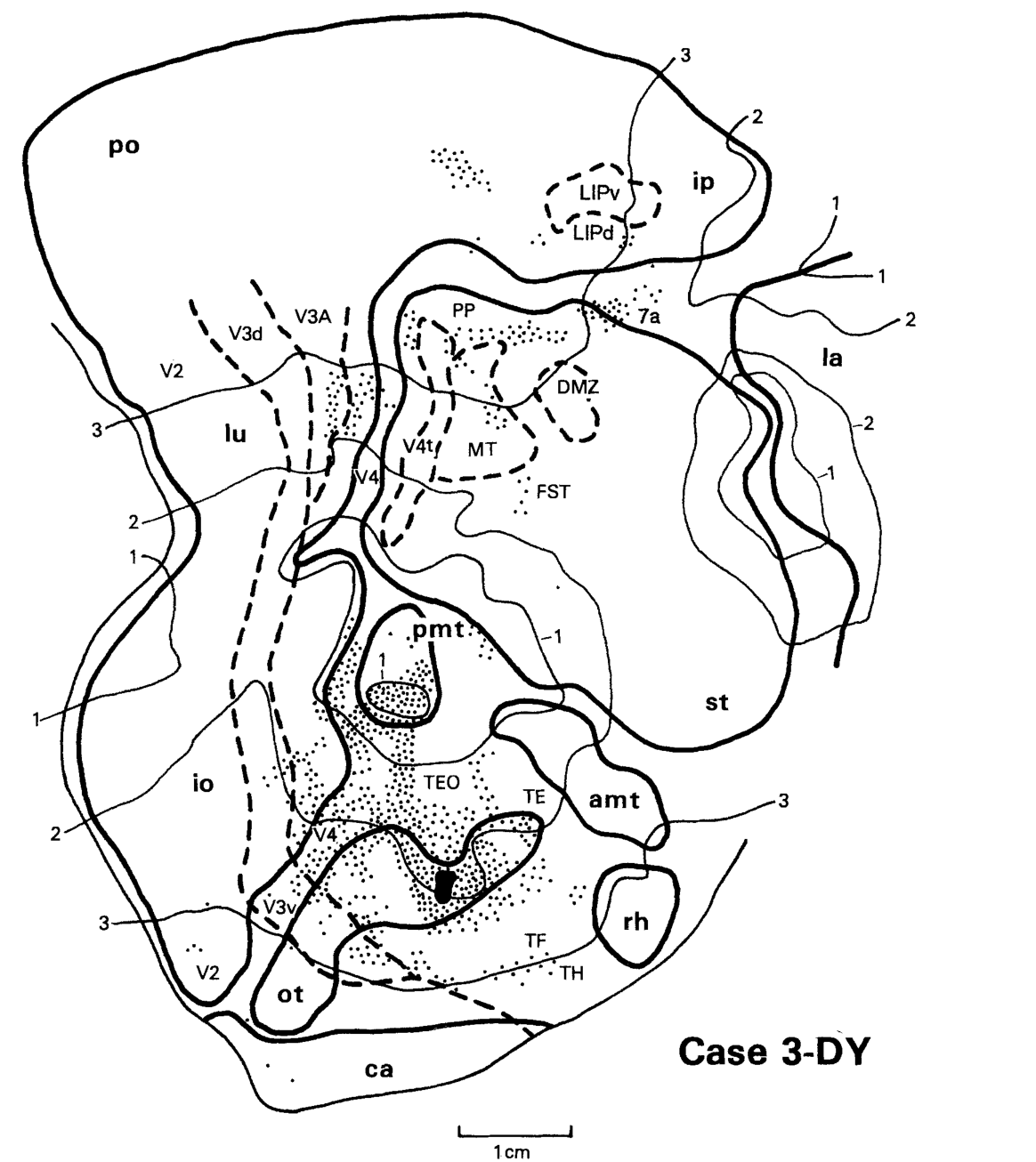
图 10 · 上视野代表区的顺向终末标记
原文图注:Fig. 10. Case 4-AA: Location of anterogradely labeled terminals after a tritiated amino acid (AA) injection into the peripheral upper field representation of TEO. Conventions are as in Figures 1-3 and 5.
氚标记氨基酸注入上视野代表区后的顺向终末标记分布:轴突终末横跨背侧 V3A/V4 与腹侧 V3v/V4 垂直子午线代表区,PP 区的终末呈全层均匀分布,符合中间型连接的判定标准。
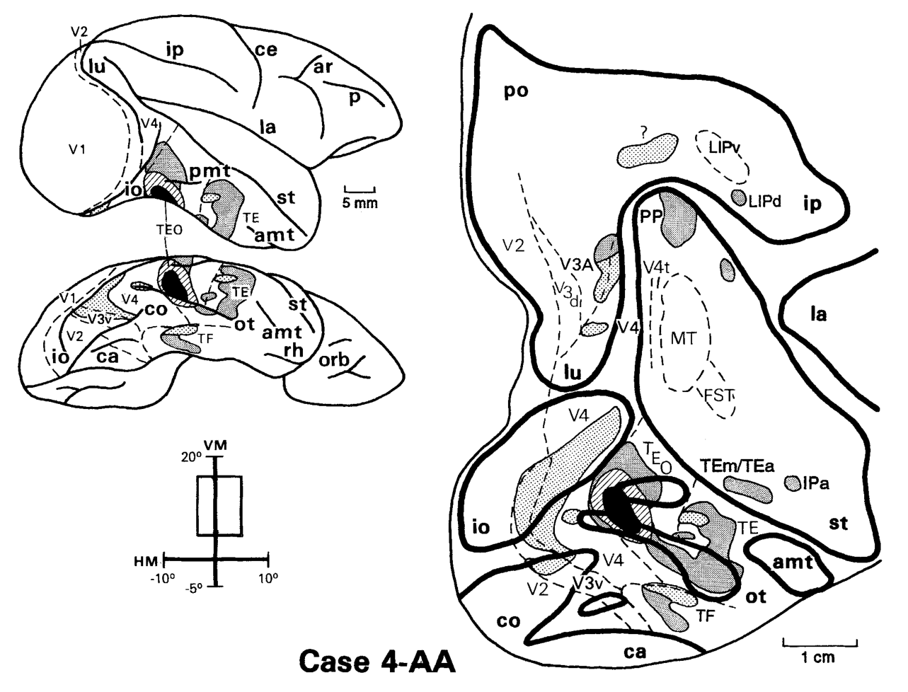
图 11 · 下视野代表区注射(DY)的逆向标记
原文图注:Fig. 11. Case 1-DY: Distribution of retrogradely labeled cells after a DY injection into the peripheral lower field representation of TEO. Arrow (upper left) points to the location of the injection site buried in the sulcus. Conventions are as in Figures 1-3.
下视野周边代表区(偏心度 26°)注入 DY 后的逆向标记:标记细胞系统性出现在 V2、V3、V4 的背侧(下视野)区域,吻侧主要标记 TE、TF、TH 与 36 区,与图 8(上视野)构成空间镜像互补。
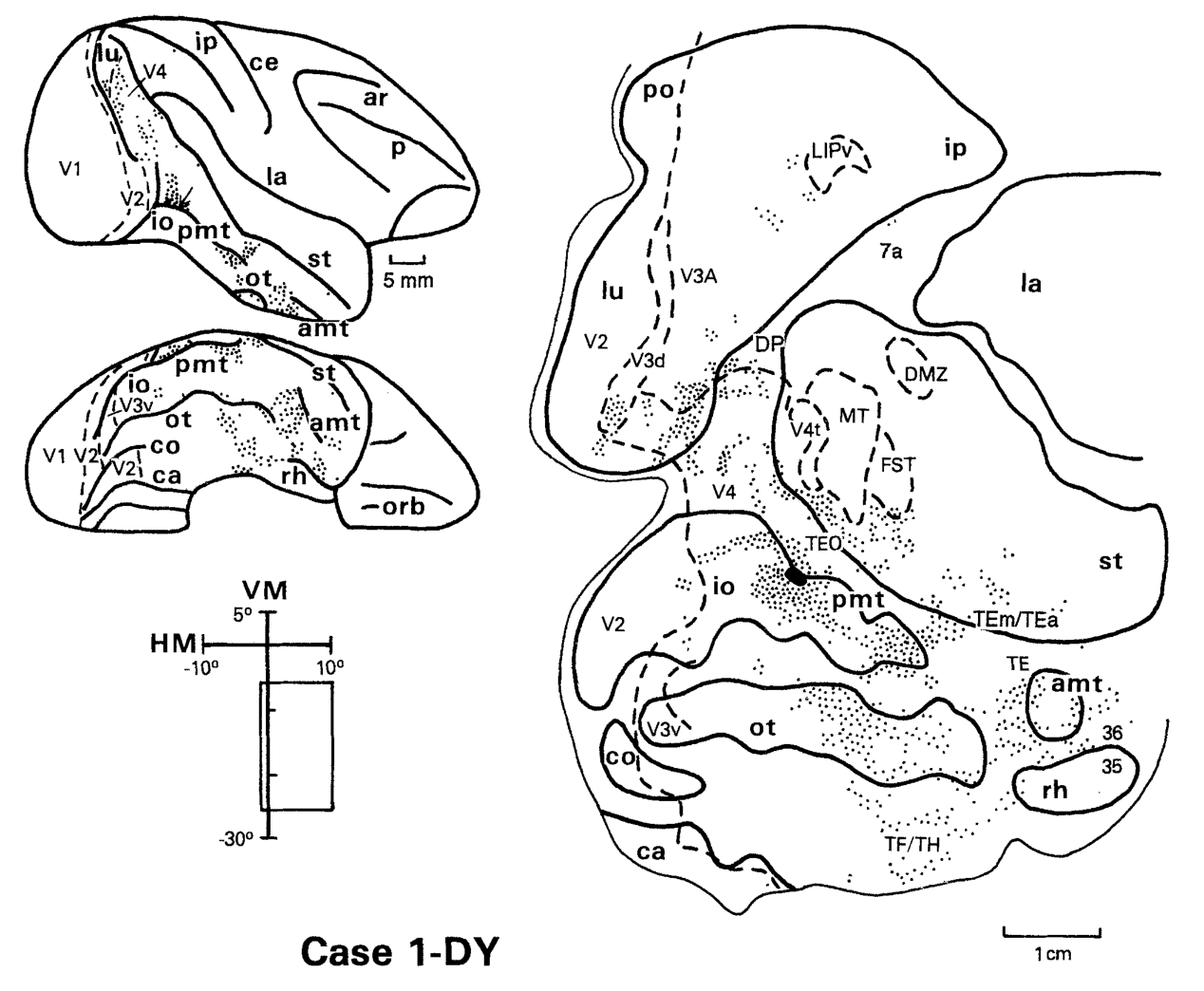
图 12 · 下视野代表区的顺向终末标记
原文图注:Fig. 12. Case 1-AA: Distribution of anterogradely labeled terminals after an AA injection into the peripheral lower field representation of TEO. Conventions are as in Figures 1-3 and 5.
下视野代表区顺向示踪切片分布:终末在背侧 V2、V3d、V3A、V4 避开第 IV 层(反馈),在 TE 与 TEm/TEa 集中于第 IV 层(前馈),在 LIP 与 IPa 呈全层分布(中间型),在一例个案中完整呈现三种连接层型。
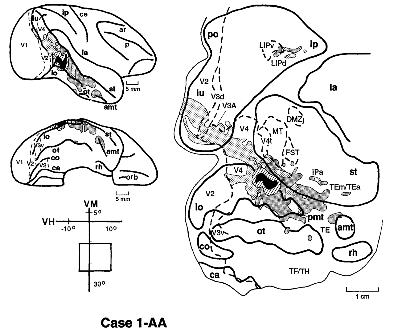
图 13 · 意外打入 PITd 的对照例案
原文图注:Fig. 13. Case 2-FB: Distribution of retrogradely labeled cells after an FB injection into PITd. Arrow (top) points to the location of the injection site buried in the sulcus. Conventions are as in Figures 1-3.
误注入相邻脑区 PITd 的对照个案逆向标记图:整体标记与 TEO 例案存在相似性,但早期视皮层 V2 与 V3 区域完全缺失细胞标记,为区分 TEO 与 PITd 提供了明确的连接组学标准。
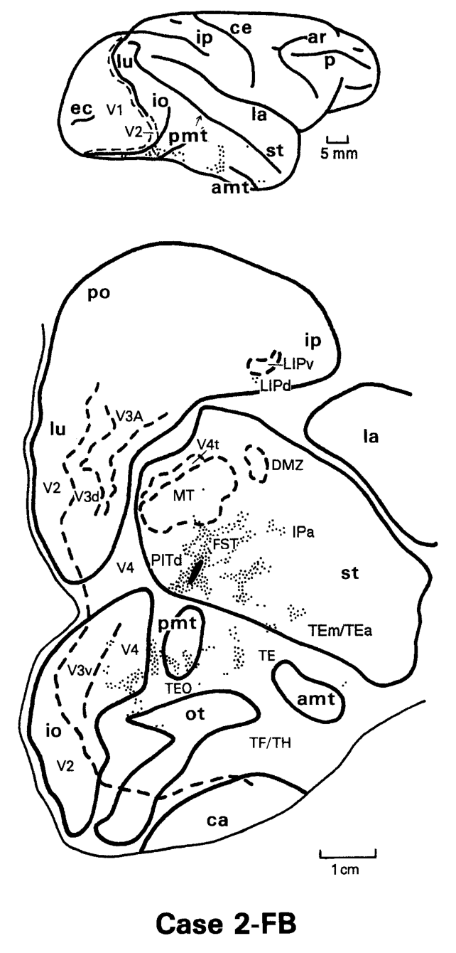
图 14 · 额叶眼区(FEF)的标记
原文图注:Fig. 14. Location of retrogradely labeled cells in the frontal lobe after foveal (case 1-WGAr) and peripheral (case 2-WGAr) TEO injections. The positions of the sections shown on the bottom are indicated on the lateral (case 1-WGAr) and on the dorsal views of the brain (case 2-WGAr), respectively. Arrows (top) point to the regions within the arcuate sulcus where labeled cells were found. Conventions are as in Figure 2.
对比中央凹注射与周边视野注射在额叶引起的标记分布:标记神经元均聚集在弓状沟下支前岸的额叶眼区(FEF)内,未表现出明显的视网膜拓扑空间位移,表明 TEO-FEF 连接主要参与非拓扑组织的眼动调控通路。
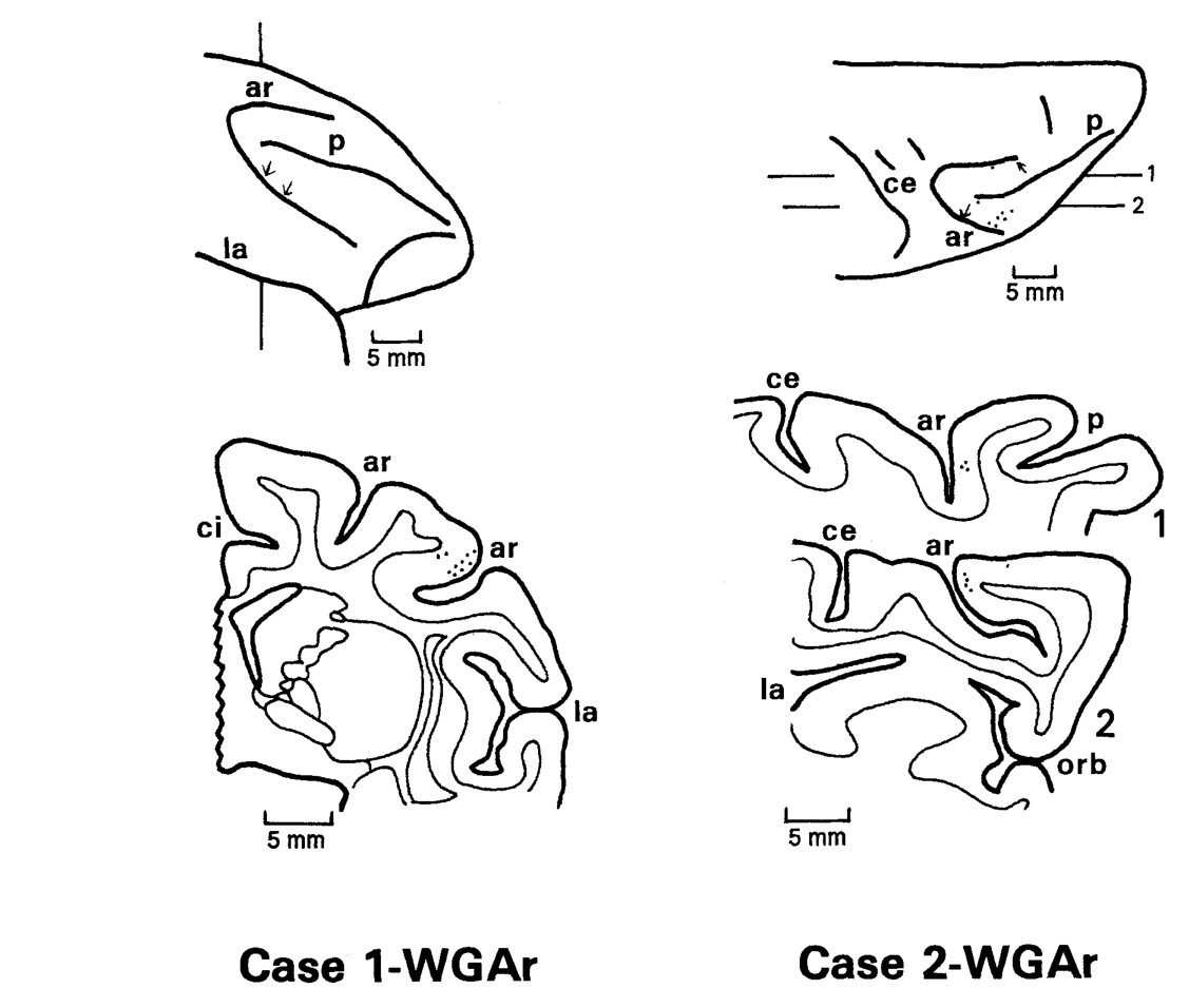
图 15 · TEO 连接总示意图(箭头=层型)
原文图注:Fig. 15. Schematic illustration of the connections of area TEO, shown on a lateral view of the macaque brain. The lunate, intraparietal, superior temporal, and arcuate sulci have been opened up to reveal the location of visual areas within them. Solid arrowheads indicate feedforward connections, i.e., those in which labeled terminals are heaviest in layer IV; open arrowheads indicate feedback connections, i.e., those in which labeled terminals are lightest in layer IV, and reciprocal solid arrowheads indicate intermediate-type connections, i.e., those in which labeled terminals are evenly distributed across all layers. ……
在脑侧面展开示意图上总结 TEO 的综合皮层连接模式:实心箭头表示前馈连接(终末以第 IV 层为主),空心箭头表示反馈连接(终末避开第 IV 层),双向箭头表示中间型连接(全层均匀分布),系统呈现了 TEO 的投射极性。
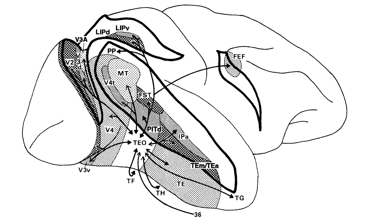
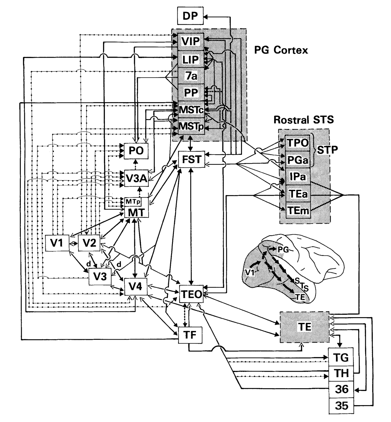
图 16 · 视皮层层级总图与 TEO 的位置
原文图注:Fig. 16. Summary diagram of the visual cortical hierarchy (adapted and updated from Ungerleider and Desimone, '86b; Boussaoud et al., '90). Solid lines indicate connections originating from both central and peripheral field representations, while dotted lines indicate connections restricted to peripheral field representations. Solid arrowheads indicate feedforward connections, open arrowheads feedback connections, and reciprocal solid arrowheads intermediate-type connections (see figure 15). The diagram demonstrates the crucial anatomical position of TEO within the inferior temporal (object vision) processing stream as well as its potential for communication with the parietal (spatial vision) and superior temporal (motion analysis) processing streams. ……
灵长类视皮层层级连接综合框架图:实线代表全视野共同连接,虚线代表仅限周边视野代表区的投射回路。展示了 TEO 位于枕颞物体识别通路中的枢纽地位,以及通过中间型连接与顶叶(空间视觉)和上颞沟(运动加工)保持双向交叉交互的解剖结构。
讨论
作者从三个维度评估了 TEO 的连接组织原则:
首先,在层级分类方面,依据严格的层板终止特征,V2、V3、V4 及 MT 向 TEO 提供前馈输入,并接受来自 TEO 的反馈投射;TEO 向 TE、TEm/TEa 与 IPa 发出前馈投射,并接受其反馈调控;而 TEO 与顶叶(LIP、PP)、额叶眼区(FEF)、STS 区域(FST、PITd)及海马旁回 TF 区表现为相互的中间型连接。作者指出,将 TF 置于与 TEO 相当的中间层级,反映了边缘系统与高阶视皮层连接的复杂性。
其次,在功能分工方面,TEO 位于腹侧识别通路的中心枢纽,其接受 V4 的密集输入并主要输出至 TE,该解剖层级与其介于局部特征编码与复杂物体表征之间的生理调谐特性相吻合;损毁研究中 TEO 切除损害二维图形辨别而保留记忆、TE 切除主要损害再认记忆的现象,获得了直接的解剖回路支持。
最后,针对既往研究中“损毁 V4 后 TE 神经元仍保有视觉反应”的现象,本研究证实 V2 与 V3 存在绕过 V4 直接进入 TEO 的投射通路,且文献表明投向 TEO 与投向 V4 的 V2 神经元处于相同的细胞构筑条带中,表明腹侧通路内部存在平行的前馈旁路通路。此外,TEO 周边视野代表区特异性地与背侧空间及运动视区保持双向连接,提示高阶腹侧皮层在视野外周保留了空间定位与运动检测的交互界面。
一句话总结
本研究通过电生理定位与层板双向示踪技术,阐明了猕猴 TEO 皮层作为腹侧物体识别通路关键前馈中继站的解剖连接网络,揭示了 V2/V3 绕过 V4 直达 TEO 的旁路结构,并证实周边视野代表区特异性地与背侧运动和空间通路保持多重中间型连接。