这是一篇科学史考证型综述,作者把猕猴枕颞交界区 TEO 的"出生证明"从头查了一遍。核心发现是:TEO 这个名字虽然出自 Bonin 与 Bailey 1947 年的猕猴细胞构筑学图谱,但那份图谱其实拿不出把 TEO 与邻近区分开的证据;真正第一次把它与 TE 区分开的是 1949 年的马钱子碱神经元描记(连接证据),1969 年 Iwai 与 Mishkin 的皮质切除-行为实验又补上了因果证据。这篇文章值得读,一方面它纠正了"TEO 由细胞构筑学定义"这一流传甚广的误会,另一方面它找回了 1970 年代就已使用、却常被记到 1990 年头上的 PIT/AIT 命名史。
研究背景
脑区的名字远比表面看起来混乱。Gross(1994)早在二十多年前就把各种脑区缩写比作"字母汤"(alphabet soup),而随着测量与绘图手段增多,这锅汤只会越来越稠。同一块皮层,可以按细胞构筑学(cytoarchitecture,基于染色切片分层特征)、按连接、按功能分别定义,各有各的标签。TEO 位于猕猴枕叶与颞叶交界处,是腹侧视觉通路(ventral stream,物体识别通路)的重要一段,现代文献普遍把它的定义归功于 Bonin 与 Bailey(1947)。但作者在与众多视觉认知研究者的交流中发现,大家都默认 TEO 是"temporal(TE)+ occipital(O)"的合成词——而事实上当年没有任何一篇论文写明 TEO 究竟代表什么,这个名字的来源本身就是一段失考的历史。
当时通行的脑图体系有两套:Brodmann(1907)的数字命名,把这块过渡区叫作 area 37;Economo 与 Koskinas(1925)的符号命名,把它叫作 PH。Bonin 与 Bailey 在专著开篇明确声明弃用 Brodmann 的数字系统、改用 Economo 的符号——理由是他们最终目标是理解人脑,而 Brodmann 人脑分区的原始数据从未发表,Economo 与 Koskinas 的图谱细节完整、便于比较。缺口在于:没人认真核查过 Bonin 与 Bailey 是否真的有细胞构筑学证据支持 TEO 是一个独立脑区,而与 TEO 定义关系密切的 Iwai 系列研究大多是会议论文集,"躺在图书馆书架上",基本被现代文献遗忘。作者认为,保存这段证据史和时间线,是防止定义与命名张冠李戴的前提。
研究思路
作者的总体策略是逐篇核查原始文献、大段引用当事人原话、按时间线重建证据链。全文七节依次推进:TEO 名字的来源 → Bonin 与 Bailey 在细胞构筑学上的困境 → 马钱子碱神经元描记(strychnine neuronography)如何首次区分 TEO 与 TE → Iwai 与 Mishkin(1969)的切除-行为证据如何确立 TEO 的后边界 → 1970 年代 PIT/AIT 命名的兴起与 HRP 连接证据 → 1990 年 PIT 命名重现及其现代对应 → TEO 功能异质性与命名方法的现代讨论。作者在脚注里交代了取舍:他聚焦切除与神经元描记研究,因为它们在历史上对 TE、TEO、OA 的区分起决定作用;同时期 Gross 等人的电生理研究超出本文范围。这种"让史料自己说话"的写法,是为了把每类证据的贡献精确归位,而不是替历史下裁决。
方法
这不是一篇实验论文。作者在文末伦理声明部分明确写道,本文是历史综述,没有开展任何人类或动物实验。材料是 1943–1990 年间的原始论文、图版与图谱:Bonin 与 Bailey(1947)猕猴专著的 Plate L 及其示意图(Section 800、840–890 的切片图版)、Petr 等(1949)的马钱子碱神经元描记记录、Iwai 与 Mishkin(1969)发表于 Experimental Neurology 的切除-行为实验、Iwai(1978、1980、1982)与 Iwai 等(1990)的会议论文集与期刊论文、Umitsu 与 Iwai(1980)的辣根过氧化物酶(horseradish peroxidase, HRP)追踪实验。方法的核心是把不同年代的图版、图注与正文表述相互对照,重建"谁在什么时候用哪种证据说了什么"。
对不熟悉这几个历史手段的读者,可以先明确它们各自说明什么:细胞构筑学靠染色切片的分层形态划分脑区,是"结构"证据;马钱子碱神经元描记术把马钱子碱置于一个皮层位点,其诱发的电活动沿轴突传导到相连皮层,借此描绘皮层间连接,是"连接"证据;皮质切除(cortical ablation)通过移除皮层观察行为缺损,是"因果功能"证据;HRP 追踪则是向目标区注射示踪剂、标记与之相连神经元的现代解剖连接手段。这四类证据在文中的先后登场,正好构成了本文的叙事骨架。
主要结果
-
TEO 的名字是"借来的",而且 Bonin 与 Bailey 自己也不确信它是独立脑区(图1、图2、图3a)。Economo 与 Koskinas 的符号命名规则是:大写字母代表脑叶、花体字母代表回、下标代表显微特征,当区域跨越脑叶时再加第三个大写字母——顶叶第 8 区 PH 延入颞叶的部分记作 PHT、延入枕叶的部分记作 PHO。既然 Bonin 与 Bailey 模仿这套命名,TEO 最可能就是按同样逻辑表示"TE 延入枕叶的部分"。但在整个 1947 年专著里,TEO 只在 Plate L 的标题中出现一次:它被排除在图谱正页(frontispiece)之外,示意切图(Section 800)里 OA 与 TE 之间的那块皮层被标成"?"而非 TEO——他们自己都没把握这是细胞构筑学上可分的区域。
-
第一个真正把 TEO 与 TE 区分开的证据是连接,而非细胞构筑(Petr 等,1949)。将马钱子碱置于第三颞回后部(即 TEO 对应区)时,它向第二颞回、梭状回、月状沟前后(OA 与 OB)以及顶内沟上下(PEp 与 PG)放电,还能从主沟(sulcus principalis)与角回接受逆向放电。Petr 等明言:"虽然 Bonin 与 Bailey 未能把它认作独立的细胞构筑区,但它具有这样的特征性连接,使人几乎不容怀疑,它是 Economo 在人类所称的可分区 PH 的前身"。也就是说,TEO 与 TE 在连接谱上不同——这是该区第一次被明确区分。
-
切除-行为实验提供了因果证据,并确定 TEO 的后边界(Iwai 与 Mishkin,1969;图3b)。切除实验显示 TEO 与 TE 引起不同的行为缺损,二者功能可分;后续实验(Iwai,1980;Kikuchi 与 Iwai,1980)进一步确定 TEO 的后界在枕下沟升支(ascending limb of the inferior occipital sulcus, AIOS)——是否包含 AIOS 对图形辨别行为的损害至关重要。Iwai(1980)总结道:视觉学习区由 TE(AIT 亚区)与可能的 TEO(PIT 亚区)组成,不延伸到 OA 与 OB。
-
PIT/AIT 命名其实在 1970 年代就已通行,比通常的归属早了十多年(图4)。Wilson 等(1972)、Iwai(1978)、Sahgal 与 Iversen(1978)、Bolster 与 Crowne(1979)等切除研究已使用 posterior inferotemporal(PIT)/anterior inferotemporal(AIT),而现代文献常把这对命名归于 Van Essen 等(1990)。同期 Cowey 与 Gross(1970)、Gross(1973)主张"foveal prestriate"更贴切,Iwai(1978)则反驳说 foveal prestriate"至少是不恰当的",并正式提议用 AIT、PIT 指代视觉学习区的前、后亚区。Iwai 的处理是双标签并行:解剖学定义用 TE/TEO,功能(切除)定义用 AIT/PIT。
-
TEO 在解剖连接上独立,且与顶叶有垂直连接——"形状—空间"交互模型的先声(图5)。Umitsu 与 Iwai(1980)用 HRP 证明后下颞皮层与前下颞皮层、纹前皮层在解剖连接上可分,并且垂直连向顶内沟(intraparietal sulcus, IPS),与 Petr 等(1949)的发现一致。两年后 Iwai(1982)据此提出"形状视觉系统"与"空间视觉系统"可经这些垂直连接"相互交互"。这与 Ungerleider 与 Mishkin(1982)提出两皮质视觉系统假说几乎同期,而"垂直连接承担两通路间串话"的角色直到 Takemura 等(2016、2017)才被重新提出——作者认为 Iwai 的提议相当前瞻。
-
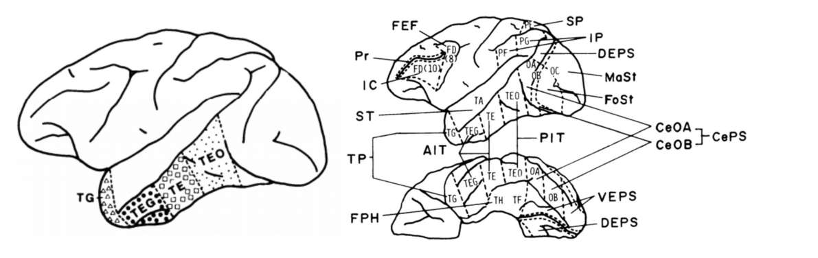
同一篇论文里两套标签并存的做法至今仍在延续(Iwai 等,1990;图6)。就在 Van Essen 等(1990)重新提出 PIT 命名的同一年,Iwai 等(1990)在同一篇论文的解剖部分用 TEO/TE、在行为切除部分用 PIT/AIT。Van Essen 等(1990)引用 Iwai 时之所以取 TEO 而非 PIT,正是因为所引工作属于解剖性质——这恰好印证了 Iwai"按方法选标签"的规则。
图注解读
图 1 · "TEO"标签的由来:从 PH 到 TEO 的类比
原文图注:Fig. 1 The origin of the "TEO" label. As specified in the Introduction of the main text, Bonin and Bailey admired the work of Economo and Koskinas and preferred their nomenclature over Brodmann's nomenclature. Nevertheless, Economo and Koskinas (1925) identified a transitional area ("PH") between areas OA and TE in human (a left) that Bonin and Bailey (1947) could not identify in macaque (b right). As Economo and Koskinas also identified a portion of PH extending into the occipital lobe, which they labeled PHO (Fig. 1a, top), it is likely that Bonin and Bailey mirrored the logic of Economo and Koskinas to generate the "TEO" label to represent the portion of area TE extending into the occipital lobe. However, note that TEO is excluded from the frontispiece of their monograph.
这张图左右并排对比两套脑图:a 是 Economo 与 Koskinas(1925)的人脑分区图,外侧面上用红色虚线圈出 PH、PHT、PHP、PHO,内侧面显示 PH 向腹侧颞叶延伸;b 是 Bonin 与 Bailey(1947)的猕猴脑图,外侧与腹侧面里 OA 和 TE 之间都没有标出 TEO。要点是"有 PH 的体系"与"没有 TEO 的图谱"形成对照:人类有人类专属的过渡区 PH 及其跨叶变体 PHO,猕猴图谱却没有对应的独立过渡区。这直接支撑结果 1——TEO 这个名字很可能是按 Economo 的跨叶加字母逻辑类推出来的,而它连自己的图谱正页都进不去,说明命名先于证据。
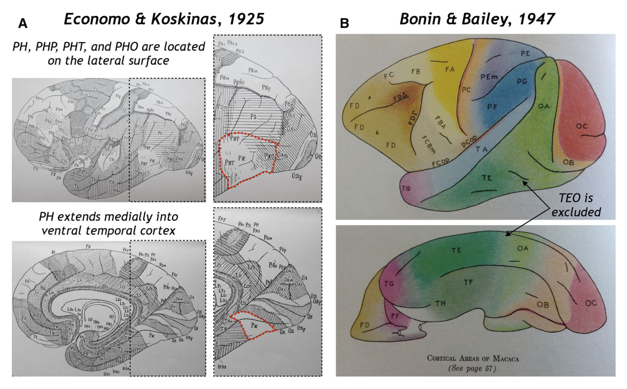
图 2 · 1947 年图版里的 TEO:与 OA、TE 长得一样
原文图注:Fig. 2 Bonin and Bailey (1947) had difficulty differentiating area TEO from neighboring areas TE and OA based on cytoarchitecture. Plates from Bonin and Bailey (1947) illustrating the cytoarchitecture of TE, TEO, and OA between Sections 840 and 890. While TEO was not included in their frontispiece as depicted in Fig. 1b, Bonin and Bailey included TEO in their plates to show the cytoarchitectonic similarity with areas OA and TE (a direct account of their description is included within the section "What is the origin of the "TEO" label?" of the main text)
改为 '细胞构筑学染色切片图版' 或删去 '尼氏' 二字;若保留需注明为解读者的补充而非原文信息。读图的关键是:把 TEO 的图版与左右邻区的图版并排看,分层形态彼此相似,看不出可以划界的差别——这正是原文引用的困惑("Plate L 取自该中间区域,第五层很浅,与 OA 相似")。它支撑结果 1 的第二半:TEO 不仅没进正页图,连他们放出的图版本身也只是用来呈现它与 OA、TE 的相似,而非差异。
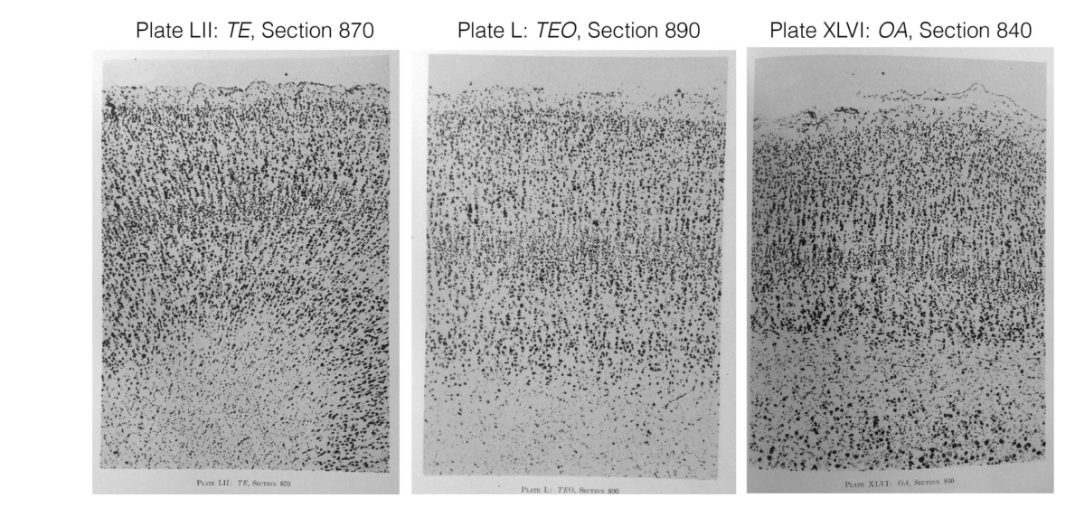
图 3 · 1947 年的"?"与 1969 年的重建
原文图注:Fig. 3 Area "?" in 1947 and re-created over two decades later. a Figure 15 from Bonin and Bailey (1947) depicting a schematic illustration of Section 800. Note that the piece of cortex between OA and TE is labeled with "?" and not "TEO." This is because they were unsure that the piece of cortex between OA and TE was different from either area based on cytoarchitecture. b Iwai and Mishkin (1969) re-created the location of slice 800 to relate Bonin and Bailey's cytoarchitectonic confusion to the location of their ablation studies. They concluded that while TEO is hard to cytoarchitectonically differentiate from TE, behavioral evidence after cortical ablation indicates that these are indeed separable areas (details of which are included in the section "Cortical ablation studies functionally differentiate TEO from TE and solidify the posterior boundary of TEO at the ascending limb of the inferior occipital sulcus" of the main text). The original caption from Fig. 4 of Iwai and Mishkin (1969) reads: "Cytoarchitectural division of the occipito-temporal region after Bonin and Bailey (3). Top, line drawing based on original colored map. Middle, section No. 800 showing unidentified ("?") area. Bottom, approximate reconstruction of unidentified area." (Iwai and Mishkin 1969, pg. 592; note that the original images were re-organized to accommodate the organization of the present figure)
这张图分上下两半讲同一条皮层在两个年代的身份。上半(a)是 Bonin 与 Bailey 的 Section 800 示意图,OA 与 TE 之间那块皮层明晃晃地写着"?";下半(b)是 Iwai 与 Mishkin(1969)按原彩色图重绘的线图,把这块"身份未定"皮层的位置重建出来,并与他们的切除位点对齐。解读时要注意时间差:1947 年标"?"是因为细胞构筑学分不清,1969 年的重建则是为了给行为证据定位——同一块皮层,先有"?"后有"可分"。它支撑结果 1 与结果 3 的衔接。
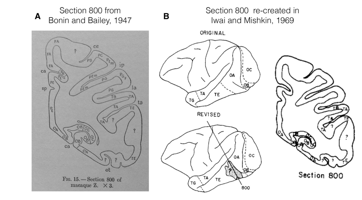
图 4 · 同一块皮层、两套标签:Iwai 的双命名策略
原文图注:Fig. 4 PIT, PH, and TEO in macaque by Iwai (1978, 1980). Top: Fig. 1 from Iwai (1978) in which he used PIT and AIT to refer to areas TEO (or PH) and TE, respectively. As described in the main text, Iwai used the TEO and TE nomenclature to refer to areas defined anatomically and used the PIT and AIT nomenclature when referring to areas defined functionally in his ablation studies with Mishkin and others. This is why there are two labels in each image. The original caption reads: "Two visual learning foci in the temporal association cortex of monkeys, and diagrammatical representations of various cortical areas. The visual learning area consists of two anatomically and functionally distinguishable subareas. The anterior inferotemporal subarea (AIT) or Area TE (dotted area) is concerned w……"(后略,原注继续说明 AIT/TE 负责视觉记忆或联结学习、PIT/PH 或 TEO 负责图形知觉与选择性注意,foveal prestriate 皮层在视觉学习区之外)
这张图是 Iwai(1978,上)与 Iwai(1980,下)两张猕猴脑示意图,每张图上同一块皮层都挂着两个名字:脑图本体上是解剖学标签 TE 与 PH(或 TEO),括号在外的是功能标签 AIT 与 PIT。读图时先分清内外两圈标签的对应关系——AIT=TE、PIT=TEO(或 PH)——再看两组功能描述的差异:前亚区管视觉记忆与联结学习,后亚区管图形知觉与视觉辨别学习中的选择性注意。它支撑结果 4:PIT/AIT 命名在 1970 年代已被切除研究者使用,且 Iwai 明确区分"解剖定义"与"功能定义"两套称呼。
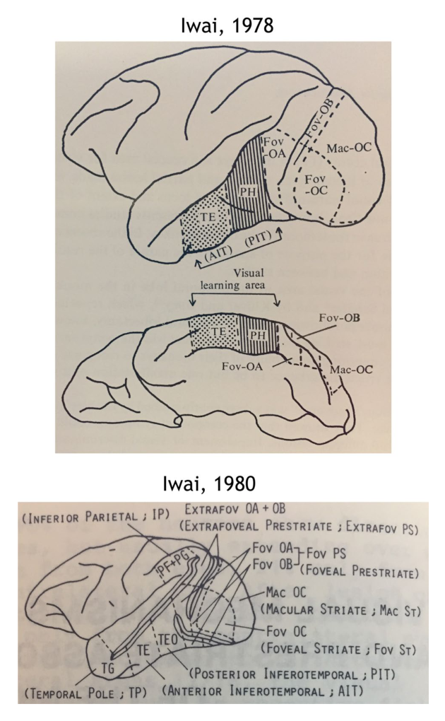
图 5 · TEO 的解剖连接:垂直连接可能整合形状与空间两条系统
原文图注:Fig. 5 Anatomical connectivity of TEO: vertical connections may allow integration between "form vision system" and "spatial vision system." Umitsu and Iwai (1980) used horseradish peroxidase (HRP) to examine if the posterior inferotemporal cortex was distinguishable from adjacent pieces of cortex based on anatomical connections. a Original caption reads: "Two representative cases in which multiple HRP injections were made into the posterior inferotemporal cortex (area TEO, Fig. 1a) and into the lateral prestriate cortex (Fig. 1b). Hatched areas: HRP injection sites. Filled circles: HRP-labeled cells in the supgragranular layers. Open squares: HRP-labeled cells in the……"(后略,原注说明大符号代表 100 个标记细胞、小符号代表 10 个;b 为 Iwai 1982 年提出的视觉信息加工模型)
这张图 a 部分是两例 HRP 注射的平铺图:分别向后下颞皮层(TEO)与外侧纹前皮层多点注射,阴影为注射区,实心圆与空心方块分别标记上层与下层中被标记的细胞,符号大小对应标记细胞数量级。读图重点在标记细胞的分布位置:TEO 注射逆行标记出的细胞分布格局与纹前皮层注射不同,说明 TEO 在连接上自成一区,尤其连向顶内沟——与 Petr 等(1949)的垂直连接证据遥相呼应。b 部分是 Iwai(1982)的四系统模型示意,从 IPS 出发的垂直箭头表示"形状视觉系统"与"空间视觉系统"经此交互。它支撑结果 5。
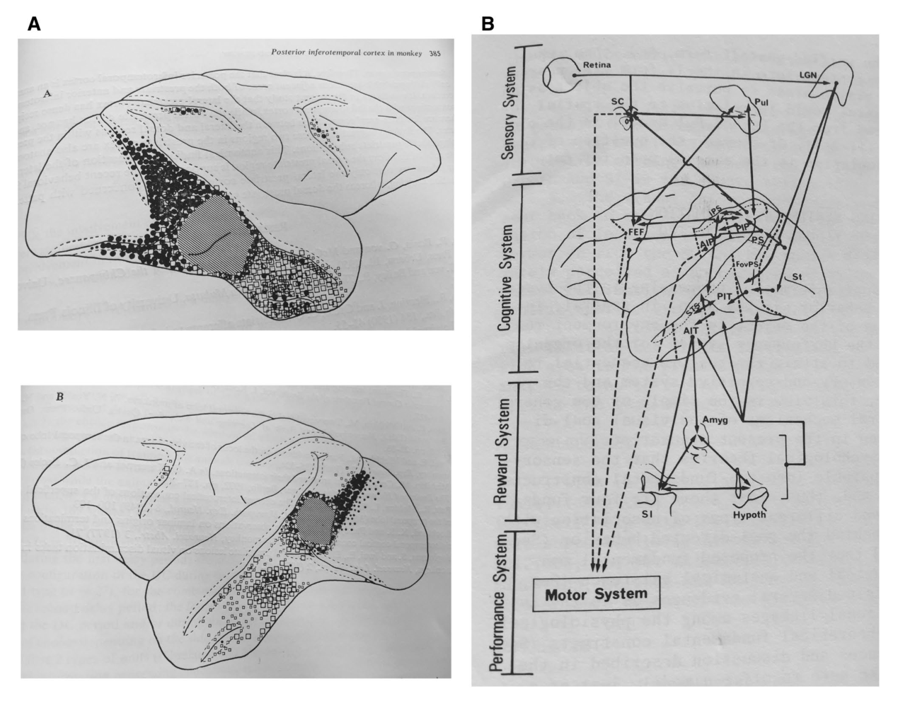
图 6 · 同一篇论文里的 TEO 与 PIT:方法决定标签
原文图注:Fig. 6 TEO and PIT: Multiple labels to refer to the same piece of cortex depending on the methodology in the same study. The same year that Van Essen et al. (1990) re-proposed the PIT nomenclature, Iwai et al. (1990) used both the TEO and PIT nomenclature in the same paper in which TEO was used during the first anatomical portion of the study (left) and PIT was used during the later behavioral and ablation portion of the study (right). The fact that Iwai used multiple labels to refer to the same piece of cortex depending on the methodology is an approach that is still used today—for example, using separate labels to refer to cortical areas if they are either defined based on cytoarchitectonic staining or functional magnetic resonance imaging (see the sections "PIT and AIT re-emerge in 1990"……)(后略)
这张图左右两幅都出自 Iwai 等(1990)同一篇论文:左图是解剖部分,图注原文为"TG、TEG、TE 与 TEO 各以空心三角、大实心圆点、空心方块与小圆点示意划分";右图是行为与切除部分,用的是 AIT、PIT、FoSt、IP 等一长串功能—定位缩写。读图的关键是认出左右两图覆盖的是同一块皮层、却换了整套词汇——这不是笔误,而是"解剖研究用 TEO/TE、功能研究用 PIT/AIT"规则在同一篇论文里的现场演示。它支撑结果 6,也点出这种双标签策略与现代"细胞构筑 vs fMRI 功能定义分开命名"的通行做法一脉相承。
讨论
作者对自己的定位很克制:他并不主张 Iwai 的做法解决了"哪个命名、哪种分区才正确"的难题——事实上同一块皮层叫 TEO、PH、37、PIT、V4A 的争议到 1990 年代依然存在,Zeki(1996)还专门撰文追问 TEO/PIT 是否与"第四视觉复合体"(V4A)完全不同。本文的目标是保存历史:把四类证据(命名类比、连接、切除、命名演变)各自出现的年代与贡献归位。讨论部分也顺势交代了现代研究怎么接这个题目:现代切除研究显示移除包含 TEO 的皮层会损害选择性注意与空间泛化(De Weerd 等,2003a、b),并改变更前部 TE 的神经反应(Bertini 等,2004;Buffalo 等,2005);fMRI 绘图在 TEO/TE 范围内发现对颜色、曲率、物体、面孔、场景、身体选择性反应的功能斑块;光遗传、药理抑制与微刺激则证明局灶失活这些斑块足以造成知觉缺陷。经典理解把 TEO 粗分为"知觉/选择性注意"、TE 为"记忆/联结学习",如今看只是颗粒度很粗的近似。局限方面,作者坦承本文不全面讨论 TEO 的现代功能证据,电生理脉络也仅在脚注提及。
与哪些文献对话之外,这篇文章给出的方法论启示同样清晰:Iwai"按方法选标签"的策略在今天依然通用——例如猕猴面孔区 PL 被不同研究描述为位于解剖学定义的 TEO 或 PITd 内,人脑的面孔选择区(pFus-faces/FFA-1)与文字选择区(pOTS-words/VWFA-1)都被发现位于细胞构筑区 FG2 之内。作者把这些例子视为 Iwai 式双命名传统的延续:不同方法定义的"同一块皮层"允许、甚至应该拥有不同的名字,前提是我们清楚每个名字背后是哪类证据。
一句话总结
依我之见,这篇考据文章最有意思的地方在于它揭示了一个反直觉的顺序:脑区命名往往走在证据前面——TEO 的名字先出现,支持它"独立成区"的连接证据和因果行为证据却分别迟到了两年和二十余年,而这段迟到的功劳簿几乎被遗忘。它提醒所有做脑区 mapping 的人:引用一个脑区标签时,最好知道自己引的到底是命名、连接还是功能的证据。
审校与证据追溯 (Verification & Evidence)
图表审计结果
- Fig1: 提取质量
good,对齐度full,识别面板[] - Fig2: 提取质量
good,对齐度full,识别面板[] - Fig3: 提取质量
good,对齐度full,识别面板[] - Fig4: 提取质量
good,对齐度full,识别面板[] - Fig5: 提取质量
good,对齐度full,识别面板[] - Fig6: 提取质量
good,对齐度full,识别面板[]
关键事实与局限性声明
- 补充要点: : (第 11 页)
- 补充要点: : (第 8 页)
- 补充要点: : (第 7 页)