一句话定位:Charles Gross 晚年对其实验室自 1965 年起猴下颞叶(inferior temporal cortex, IT)单细胞记录工作的回忆式综述。文章梳理了手与面孔选择性神经元的发现经过,系统总结了 IT 神经元的六项基本生理特性,并将电生理发现延伸至人类内侧颞叶的概念细胞及猴 fMRI 面孔斑块的研究脉络。
研究背景
下颞皮层单细胞研究的起点,是损伤实验留下的具体问题。猴下颞皮层受损后,视觉辨别学习和记忆能力显著下降,而视力等基本感觉功能及其他感觉模态的学习相对保留;这表明该区域参与物体识别,但未说明其神经元如何表征物体。与初级视觉区的感受野研究相比,下颞皮层处于视觉层级更高级阶段,其传入连接通路与反应性质当时均不明确。引入微电极记录的目的,是将区域性损毁效应推进至细胞层面,检验高级视觉区的刺激选择性特征,以及其如何整合注意调制、既往经验并实现跨视角与尺寸变化的识别。
沿用光点和光栅等简单几何图案,难以充分驱动多数下颞神经元。真实物体、手形及面孔引发的放电表明,实验需考察部件组合与整体形状配置,不能预先假定高级视觉区仅是早期视皮层滤波器的线性放大。这一方法选择最初面临解释上的质疑:复杂刺激包含多种同时变化的图像属性,研究者主观认定的手或面孔,并不等同于神经元抽象地识别了该类别概念。后续通过移除部件、重排结构、改变视角与尺寸,并在更大刺激集中测试连续反应分布,才逐渐明确其选择性特征。研究焦点由此从“是否存在面孔细胞”转向“面孔选择性反映了何种编码机制”,特别是其是否对应单一细胞与单一刺激/个体的严格对应。
这篇综述将早期记录置于后续群体编码、功能成像和因果干预的框架中进行审视。作者总结了大感受野、复杂形状偏好与相对形状恒常等特性,并根据不同细胞的选择性差异指出,偏好面孔并不等于仅响应单一特定面孔。猴功能磁共振定位的面孔斑块结合靶向单细胞记录,连接了宏观脑区与微观细胞尺度;微刺激研究则为特定细胞群参与面孔辨别提供了因果干预证据。人类内侧颞叶对特定人物照片及姓名的选择性放电提出了高度抽象表征的可能,但该性质不能直接外推至猴下颞皮层。本文属于历史性综述,早期测试集与细胞采样规模存在局限,细胞群参与特定辨别任务也不等同于独占该类知识的表征。不同皮层区域面孔细胞的功能分工、突触可塑性机制及其与临床面孔失认症的病理对应,仍有待进一步阐明。
研究思路
作者将行为学损毁证据推进至神经元水平:首先在麻醉动物中证实 IT 细胞主要对视觉刺激产生反应而非听觉刺激;随后在清醒动物任务中观察注意与记忆调制的表现;最后在固定头位条件下系统描绘其感受野属性。在刺激选择上,团队使用手、面孔及日常物体等真实图形作为测试刺激。在发现面孔与手选择性细胞后,论证核心转向检验这些细胞是否符合“祖母细胞”假设。作者分析单个细胞在 68 张图像上的连续反应分布,结合群体编码证据及人类内侧颞叶电生理记录,说明 IT 皮层主要采用群体编码而非一对一的单一细胞表征。
方法
实验对象为猕猴。早期在麻醉状态下进行单细胞记录(Gross 等 1967 年工作证实 IT 神经元对视觉刺激有特异反应);后续转入清醒固定状态绘制感受野,并在清醒行为猴上通过“延迟配对样本”(delayed match to sample)任务记录 IT 神经元在注意与工作记忆中的活动变化。刺激呈现方式包括:使用幻灯片在注视点中心投射呈现 2.5 秒,或让三维实物模型与纸剪影以约 1.2°/s 的速度横穿注视中心 15° 的窗口;刺激库中包含各种日用三维物体(Fig. 5)。数据分析主要采用刺激呈现期累积脉冲直方图(5 次试验平均)及“呈现期放电率减去前置基线放电率”的差值。综述后半部分回顾了灵长类及人类内侧颞叶单细胞记录(如 Quiroga 等 2005 年关于概念细胞的研究)以及猕猴与人类 fMRI 面孔成像实验的设计与主要发现。
主要结果
- 六条基本感受野特性(对应正文对早期工作的总结):IT 细胞有视觉感受野,但不具备视网膜拓扑定位;几乎所有感受野都包含注视中心,且对中央视野刺激反应更强;感受野范围极大(中位数约 25 平方度,部分覆盖大半视野);近半数感受野越过中线延伸入同侧视野,这在视皮层中属首次发现,并依赖胼胝体与前连合的联系;细胞偏好复杂形状或颜色,而非简单光点或光缝;对大小、对比度、颜色及位置的变化保持相对稳定的形状恒常性反应。
- 第一批“手选择性细胞”:对以注视点为中心扫过 15° 窗口的刺激进行排序,反应强度不与简单的物理维度对应,而与猴手剪影的相似程度相关;真实三维手模型诱发最强放电,去除手指或重排手指后反应减弱,连指手套式剪影反应同样偏弱(Fig. 1)。这一结果直接回应了认为记录仅反映空间频率光栅的质疑。
- 三类面孔细胞:一类对正脸反应最强,去除眼睛或嘴部后反应减弱,表现为对面孔部件的整合;一类仅对完整面孔反应,并表现出视角选择性;第三类对不同朝向均有反应,但依赖完整面孔输入。前两类反映以观察者为中心的表征,第三类反映以物体为中心的表征(Fig. 2、Fig. 3)。STP 区(前上颞沟背岸)同样记录到面孔单元,且部分细胞对生物运动敏感(Fig. 4)。
- 面孔细胞不同于祖母细胞:单个 IT 神经元对 68 张面孔与非面孔图像呈现连续分布的反应谱,对面孔反应强弱不一,对非面孔刺激也并非完全不放电(自发放电率为 20 s⁻¹)(Fig. 7)。面孔表征依托细胞群体的差异化放电模式,属于粗编码或群体编码。但在记忆系统中存在高度特异的表征:人类右前海马的一个单元对某位女演员各种装扮的照片及姓名文本均产生选择性放电(Fig. 8)。
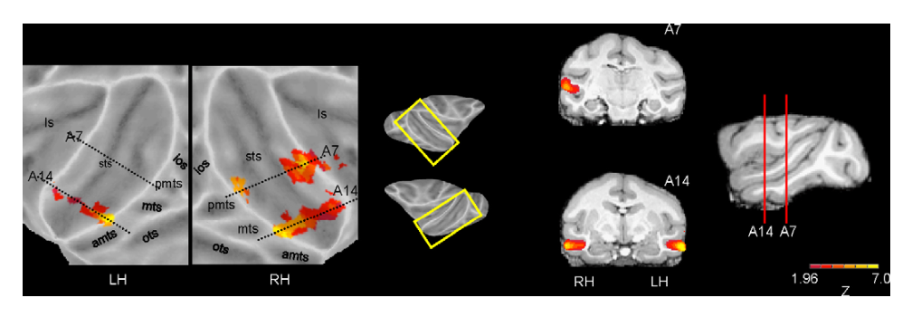
- 面孔功能簇的成像证据:猴 fMRI 显示面孔刺激在上颞沟附近诱发离散的局灶性激活(Fig. 9),与电生理中面孔细胞在 STS 附近比例达 20%–30%、其余区域约 5% 并聚集成 3–4 mm 斑块的解剖分布相符;Tsao 等(2006)在 fMRI 定位的面孔热点内记录,发现几乎所有具备视觉反应的细胞都表现出面孔选择性,功能特性类似于人类梭状回面孔区。
图注解读
图 1 · 手细胞:对"手形"的反应层级
原文图注:Fig. 1. Responses of an IT neuron that responded more strongly to hands than to any other stimulus tested. (A) Comparison of responses to hand-like and other patterns. Stimuli were a 3D model of a human hand, a cutout with the shape of the hand model, a cutout with the shape of a monkey hand, a cutout with the shape of a monkey hand with the inter-digit spaces removed ("mitten"), a partially scrambled photograph of the model of the back of the human hand, two "grating-like" hands, a 3D model of a human face and a rectangle. Stimuli were randomly interleaved and moved at 1.2°s−1 from contralateral to ipsilateral into a 15° window centered at the fovea as indicated by the horizontal lines. The histograms were based on five trials. (B) Responses to the hand cutouts in different orientations.
这张图读法:A 图展示刺激诱发的放电脉冲直方图,横线指示刺激以 1.2°/s 匀速扫过 15° 注视窗口的时程,直方图基于 5 次重复试验。反应强度排序依次为:三维人手模型 > 手形剪影 > 去除指缝的连指手套剪影 > 打乱的人手背部照片 > 条纹状手形图形 > 矩形;B 图显示改变手形剪影的平面朝向未明显削弱放电反应。这一发现表明 IT 存在偏好手形整体构型而非初级空间频率光栅的神经元。
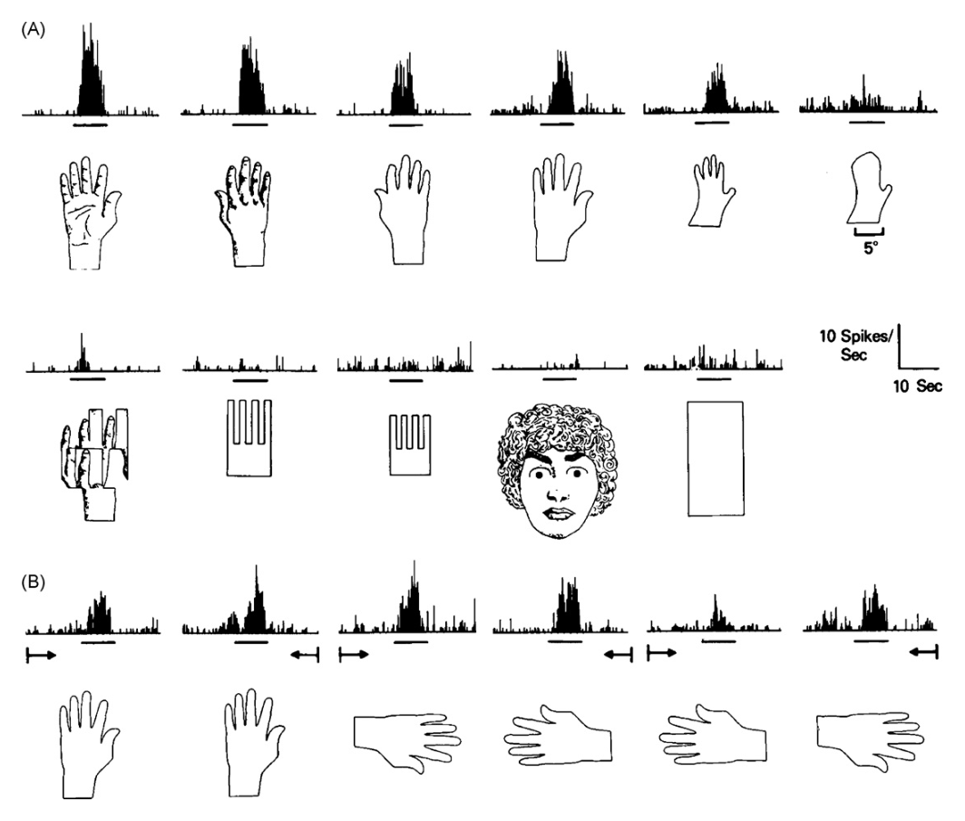
图 2 · 正脸细胞:拆掉部件反应降、朝向调反应
原文图注:Fig. 2. Responses of an IT neuron that responded more strongly to the front view of faces than to any other stimulus tested. The stimuli were interleaved colored slides projected centered on the fovea for 2.5 s indicated by the horizontal lines under each histogram. (A) Responses to a monkey face in different degrees of rotation. (B) Comparison of responses to faces, to faces with components removed and to a hand.
读法:刺激是随机交替投到注视点的彩色幻灯片,直方图下方横线指示 2.5 s 刺激呈现窗口,上方数值表示呈现期减去基线期的净放电率。A 图显示面孔旋转改变了神经元放电率,正脸激活最强;B 图显示移除眼睛或嘴部特征后反应幅度明显下降,手刺激引起的反应显著低于各类面孔条件。数据表明该细胞对面孔部件及其整体空间配置具有双重依赖。
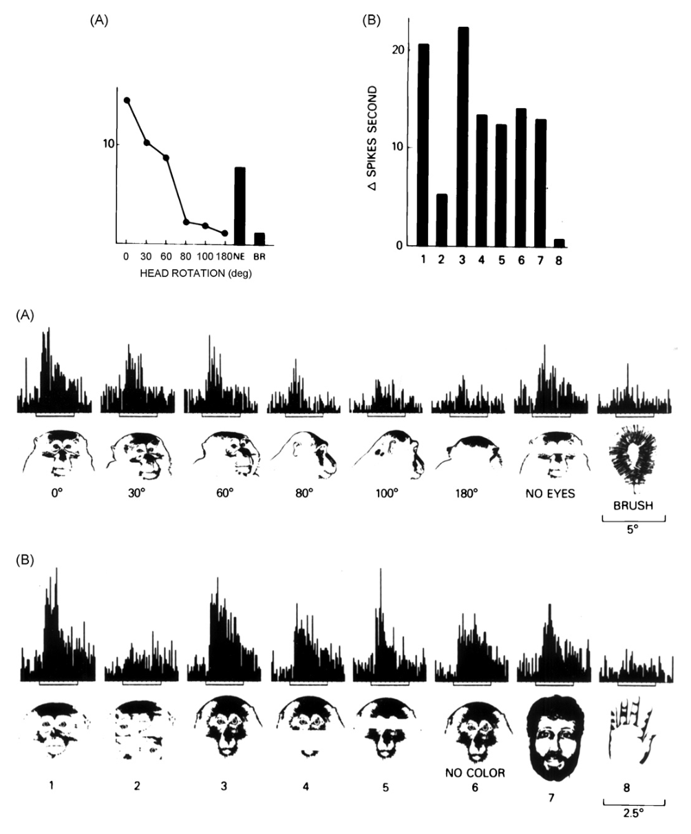
图 3 · 侧脸细胞:只认完整侧脸
原文图注:Fig. 3. Responses of an IT neuron that responded more strongly to profiles of faces than to any other stimulus tested. See also legend of Fig. 2 (after Desimone et al., 1984).
读法:实验方法与 Fig. 2 相同,此细胞的最优刺激为侧脸轮廓而非正面面孔。该结果与 Fig. 2 共同显示面孔敏感神经元群体内部存在视角选择性的亚群分化。
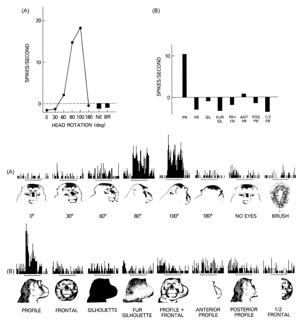
图 4 · STP 区的面孔单元
原文图注:Fig. 4. Responses of a neuron in the superior temporal polysensory area (i.e. in the dorsal bank of the anterior superior temporal sulcus) that responded better to faces than to other stimuli. Removing the eyes from the face or presenting a caricature elicited a reduced response. Cutting the face photograph into segments and rearranging the segments eliminated the response. The receptive field is shown on the lower right, C, contralateral, I, ipsilateral (Bruce et al., 1981).
读法:该记录来自前上颞沟背岸的上颞多感觉区(STP),右下图标示感受野分布(C 为对侧视野,I 为同侧视野)。人类与猴面孔诱发相似放电,刺激尺寸变化对反应影响较小;去除眼睛或使用漫画人脸后反应下降;将面孔照片分割重排后反应基本消失。该现象支持面孔整体构型是激活该类单元的关键因素。
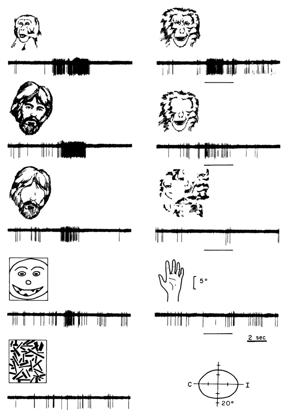
图 5 · 马桶刷细胞:影子也够用
原文图注:Fig. 5. Responses of an IT neuron that responded strongly to an oval brush. Shadows of the brush shown on the left also elicited strong responses. Presence of the handle, direction of movement and location within the receptive field (which extended 30° in all directions from the center of gaze) appeared irrelevant. Other shadows such as those shown on the right elicited weaker responses. The horizontal line indicates when the stimulus was moving through the receptive field at 8°s−1 and the numbers are the mean rate of firing during that period.
读法:左侧画出的椭圆刷及其阴影均诱发较强放电,把手存在与否、运动方向以及在感受野内的具体位置均未显著改变放电;右侧非相关图形阴影诱发的放电较弱。横线表示刺激以 8°/s 穿过感受野的区间,数值为该区间内的平均脉冲发放率。该结果展示了神经元对特定形状边界特征的抽象响应。
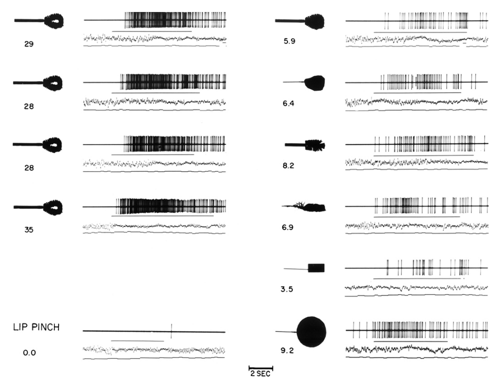
图 6 · Konorski 的"认知野"预言图
原文图注:Fig. 6. Konorski's (1967) illustrations of "particular categories of visual stimulus objects probably represented in different Gnostic fields" in inferior temporal cortex.
读法:此图引自 Konorski 1967 年著作,示意推测存在于下颞皮层中、表征特定视觉类别的“认知野”(gnostic fields),涵盖面孔、表情、躯干与日用物体等类别。作者借此说明早期实验设计选用复杂物体作为测试刺激的理论背景。
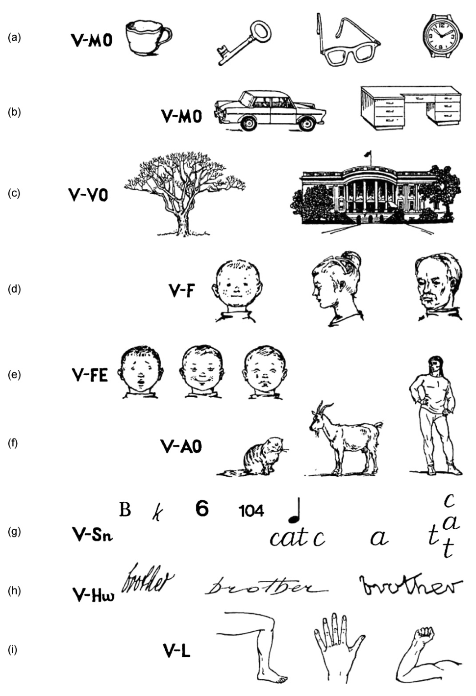
图 7 · 一个细胞对 68 张图的反应分布
原文图注:Fig. 7. Distribution of different strengths of response of one IT neuron to 68 faces and other images. F, stimuli that are front views of a face; P, face profiles; B, images that included a small face within a scene, sometimes including a whole person or other body parts. The non-face images are not labeled. The spontaneous rate of this neuron was 20 s−1 (Rolls & Tovee, 1995).
读法:横坐标按反应强度排列 68 种视觉刺激,纵坐标为放电率。F 代表正面面孔,P 代表侧脸,B 代表含有人脸的复杂场景,非面孔刺激未单独标注;基线自发放电率为 20 s⁻¹。细胞反应呈梯度分布:正脸放电最强,侧脸次之,部分非面孔物体亦引起微弱放电。这一连续响应特征为群体编码假说提供了生理学证据。
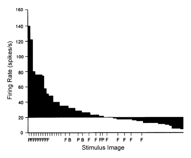
图 8 · 人类海马里的"Halle Berry 细胞"
原文图注:Fig. 8. A single unit in the human right anterior hippocampus that responds to different pictures of the actress Halle Berry including in costume and to the letter string of her name but not to other facial images or letter strings (Quiroga et al., 2005).
读法:图中展示人脑前海马记录到的单个神经元反应。该单元对特定演员的多张不同服饰与视角照片,以及该演员姓名的文字串均产生特异放电,对其他人物的面孔或名字文本则未表现出放电增加。该结果展示了内侧颞叶在概念层面的跨模态抽象表征。
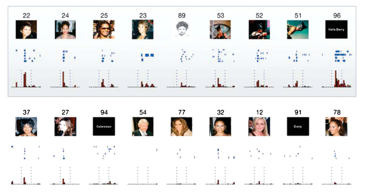
图 9 · 猴颞叶面孔功能区的 fMRI 定位
原文图注:Fig. 9. Face-selective representations in monkey temporal cortex. (Left) Flattened cortical surfaces and coronal slices in monkey M1 depicting voxels activated significantly more by faces as compared to objects. (Right) Approximate locations of the coronal slices are indicated on a sagittal slice with distances from the interaural line (mm) noted. Scale indicates Z-score values of functional activity in colored regions. RH, right hemisphere; LH, left hemisphere; ls, lateral sulcus; sts, superior temporal sulcus; ios, inferior occipital sulcus; pmts, posterior middle temporal sulcus; mts, middle temporal sulcus; amts, anterior middle temporal sulcus; ots, occipital temporal sulcus.
读法:左侧为皮层展平图与冠状切片,彩色体素展示面孔相比物体刺激激活显著增强的区域(以 Z 分数表示);右侧矢状位图标记冠状切片距双耳连线的解剖距离(毫米),并标示外侧沟与上颞沟等结构。结果显示面孔激活集中分布在上颞沟附近的离散斑块中,为猕猴颞叶的面孔功能网络构筑提供了宏观解剖学证据。
讨论
作者讨论了两个核心问题。首先是早期单细胞发现的历史发展:IT 神经元的六项基本生理特征在发表后经历多年才获得 Rolls、Perrett 以及 Richmond & Wurtz 等研究团队的系统重复与跟进。其次是表征机制的概念界定:早期关于“单一细胞表征单一概念”的严格假设与 IT 皮层的实际记录不符;IT 神经元对刺激呈现广泛而连续的反应分布,物体识别主要依托群体编码。然而,人类内侧颞叶的记录提示,在海马等记忆结构中可能存在稀疏度更高的概念表征。关于因果机制,Afraz 等(2006)在猴面孔斑块实施微电极电刺激,发现局部微刺激可显著诱导动物在辨别任务中偏向报告面孔,表明该神经元集群直接参与面孔感知的决策过程。最后,综述指出了仍待研究的问题,包括不同解剖区域(下颞凸面、STP 及杏仁核)面孔细胞的分工差异、感受野特异性的经验塑造机制,以及该神经回路在面孔失认症与自闭症中的异常改变。
一句话总结
本文系统梳理了猕猴下颞叶单细胞记录的研究历程,从基本感受野特性与复杂图形选择性的发现,阐明了物体与面孔识别的群体编码机制及其在功能磁共振成像与概念表征研究中的延续。