一句话定位:这是一篇综述,把 Rolls 团队三十余年关于灵长类(包括人类)海马"空间视觉细胞"(spatial view cells)的单细胞发现,与近年来人类 HCP 数据上的有效连接测量拼成一套完整理论:海马中的"空间"表征编码的是"你正在看哪里"(世界坐标中注视的位置),而不是"你在哪里"。它值得读,是因为它正面挑战了以啮齿类 place cell 为模板的海马叙事,并用人类连接组学证据给出空间视觉细胞如何在腹侧视觉流中"自组织生成"的机制假说。
研究背景
灵长类病灶研究早已表明,海马或穹窿损伤会损害对"外界某处"(out there)位置的学习——比如记住物体放在房间哪个位置;猴的旁海马皮层损伤甚至影响仅一对新异刺激的物体—位置联结,海马神经毒损损害空间场景记忆(Murray、Malkova、Gaffan 等的工作;人类也有对应证据,如 Crane & Milner、Smith & Milner 的右侧海马物体位置记忆缺陷)。这与啮齿类文献的主流叙事形成张力:大鼠海马研究强调的是 place cell——对"大鼠此刻所在位置"的表征,加上内嗅皮层的 grid cell,构成 O'Keefe & Nadel "认知地图"传统的路径整合与导航理论。问题在于:这套以"自身位置"为中心的框架是否适用于拥有中央凹(fovea)视觉和发达眼球运动系统的灵长类?灵长类可以不移动身体、只用眼睛扫视远方场景,它们的海马究竟表征什么坐标?
第二个缺口是连接层面。海马如何计算、表征什么,取决于它与皮层的连接,但以往灵长类空间记忆理论主要建立在解剖示踪(多来自猕猴)上,人类海马系统的输入通路——尤其是场景信息从哪些视觉区进来、自身运动信息从哪里来、奖励信息如何抵达——缺乏人类自己的量化证据。
研究思路
文章的论证策略是"细胞—计算—连接"三层递进。第一层总结猕猴单细胞记录确立的事实:海马与旁海马回的空间视觉细胞对"注视的世界坐标位置"放电,且对视网膜位置、眼位、头朝向、自身所处位置都相对不变——这需要精心设计的因子实验(同一组空间视野从不同地点、不同头朝向下观看)来排除 place、head direction、eye position 等竞争解释。第二层提出计算理论:空间视觉细胞之所以存在,是因为灵长类中央凹一次只看清约 5–10°的视野,"空间中若干特征的组合"自然定义了注视点而非自身位置;同一套竞争学习+自联想的计算过程,在大视野(>270°)的啮齿类就生成 place field,在小视野(30°)的灵长类就生成 spatial view field——这一假说用仿真检验(图 13)。第三层用人类 HCP 连接组学(172 名被试 7T fMRI,360+66 分区的有效连接测量)把细胞层面与计算层面挂到人类的解剖通路上:腹内侧视觉流提供场景"哪里",顶叶提供自身运动更新(idiothetic update),"什么"与奖励输入分别从腹外侧流和眶额皮层汇入。全文以此论证:海马在灵长类中首先是情节记忆系统,导航是记忆回溯交给新皮层(尤其顶叶)执行的产物。
方法
作为综述,方法分散在多类研究中。猕猴单细胞记录:在 2.5×2.5 m 开放测试区(位于线索丰富的大实验室内)自由觅食行走。,删去 5×5 m。,房间四壁有窗等线索;同时连续记录自身位置、头朝向和眼位(水平与垂直视线角),使每个神经元的放电都能在"位置/头朝向/注视点"三个因子上做方差分析与信息论分解。初始样本 352 个海马神经元中 40 个(11.4%)为空间视觉细胞;后续任务还包括屏幕上的物体—位置记忆、一次学习的物体—位置回忆(347 个神经元,Rolls & Xiang 2006)与奖励—空间位置联结(312 个细胞,Rolls & Xiang 2005)。虚拟现实研究(星形迷宫,Wirth 等 2017)记录眼位与 189 个海马细胞;整体位移神经元研究让猴子坐机器人做确定的旋转与平移(O'Mara 等 1994)。人类连接方面:使用 HCP 多模态分区图谱(HCP-MMP1,360 个皮层区,扩展含 66 个皮层下区),测量 172 名被试 7T fMRI 的 BOLD 信号;有效连接算法计算每对脑区在 t 与 t+tau(tau=2 s)时刻的功能连接,再经梯度下降误差修正求出方向性连接矩阵(内部用 Hopf 分岔/Stuart–Landau 振子建模),并以 Pearson 功能连接和弥散纤维束成像作交叉验证。计算理论部分配以仿真:坐标变换用增益调制(gain modulation)模型(Rolls 2020),空间表征生成用 200×200 地点网格上视场角 270°与 30°的对比仿真(de Araujo 等 2001),情节记忆存储与回溯用包含内嗅皮层、齿状回、CA3、CA1 及新皮层回投影的网络仿真(Rolls 1995 及其扩展,图 12)。
主要结果
- 空间视觉细胞的存在与坐标性质:猕猴海马与旁海马回神经元在猴子注视环境某一位置时放电,与猴子所处的位置、头朝向、面朝方向、眼位都相对无关(图 1、图 2)。信息论上,平均单细胞对空间视野的 Shannon 互信息为 0.47 bits,远高于眼位(0.017 bits)、头朝向(0.005 bits)或位置(0.033 bits);细胞自发率约 0.5 spikes/s、峰值平均 17 spikes/s(四分位距 11–20),分布在 CA1、CA3 及旁海马回。在 352 个神经元样本中 40 个(11.4%)为空间视觉细胞,其视野约占四面墙总面积的 1/16,且不同细胞视野互不相同。
- 群体编码高效且生物学上可行:关于 16 个注视位置的信息随样本神经元数(约 20 个)近似线性增长——由于信息是对数量度,这意味着群体可编码的位置数随神经元数指数增长;用最简单的线性点积解码时信息几乎不减,说明这是高维空间中的独立速率编码,而非依赖生物学上不合理的非线性解码或"噪声相关"。
- 自身运动更新:用落地窗帘遮蔽视野细节后,多数空间视觉细胞在猴子做眼动或走动转向"先前可见的注视位置"时仍继续放电几分钟,之后出现漂移,视线重现时得到校正(图 3);CA1、旁海马回、前下托在黑暗中放电只小幅下降,而 CA3 下降较大(平均降到正常反应的 23%),作者用 CA3 自联想网络的模式补全及 CA1 的直接穿通纤维输入解释这一差别。VR 星形迷宫中,28%(53/189)的海马细胞在注视地标时放电,且一些细胞在视野出现在屏幕之前、猴子把眼睛转向新位置时就放电——眼动即自身运动更新的直接证据。
- 与情节记忆的接口:开放环境中物体—位置记忆任务里,约 10% 的海马神经元对物体、13% 对空间视野、12% 对特定物体—位置组合放电;在一次学习回忆任务中(347 个神经元),以位置为回忆线索时部分神经元回忆物体、以物体为线索时部分神经元回忆位置,且回忆相关放电出现在屏幕上无任何刺激的第 4 阶段(图 4)——证明海马支持一次学会、从任一部分补全整体的物体—位置情节记忆。奖励方面,18% 的 312 个细胞对场景中偏好奖励的位置放电、5% 对次偏好奖励位置,44 个被试细胞中 60% 在奖励位置对调后几试次内翻转反应,82% 不对视觉分辨任务中的物体—奖励联结放电(奖励—空间位置而非奖励—物体)。
- 人类有效连接支持"三路输入"理论(图 5–9):腹内侧视觉流 V1>V2>V3/V4→VMV1-3→旁海马回 PHA1-3(即海马旁场景区 PSA)→海马,构成"哪里"输入;背侧流经 MT+ 复合体、顶内沟区、7 区,其中下顶叶 PGp 有强有效连接指向 PSA,被解释为对场景表征做自身运动更新所需的坐标变换(视网膜→头坐标→世界坐标方位→空间视野,图 6、7、11);腹外侧视觉流经 TF/嗅周皮层 PeEc 提供"什么"输入;眶额皮层(内侧为奖励、外侧 47m 为惩罚/非奖励)、腹内侧前额叶与前扣带回直接连接海马系统提供奖励/情绪输入(图 8、9)。另有人类特有的旁证:CA3 跨中线连接很弱,左右海马近似独立网络,右海马偏空间记忆、左海马偏语言记忆,故空间视觉细胞可能更多见于右侧海马。
- 导航理论:空间视觉细胞支持"地标到地标"(taxis)式导航——按记忆序列依次把注视转向下一个地标,不需要拓扑地图(图 10 的记忆回溯路线把空间视野信息回投到顶叶等新皮层,实现向被回忆位置的视觉运动抓取与移动,图 12);人类海马损伤只轻微影响熟悉环境中的导航、却强烈损害学习新轨迹与想象新路径,而顶叶或压后皮层损伤造成强烈的地形学失定向,这支持"海马供记忆、新皮层算导航"的分工。
- 仿真解释灵长类与啮齿类差异的同源性(图 13):视野 270°时同样的学习规则生成 place field,30°时生成 spatial view field——中央凹是两种空间表征分岔的根源;由此预测若限制大鼠视野,其海马神经元会变得更像猕猴的空间视觉细胞。
图注解读
图 1 · 空间视觉细胞 az033 的开放场地放电图
原文图注:FIGURE 1
A hippocampal spatial view cell (az033) recorded while a monkey walked around in an open field area 2.5 × 2.5 m shown as the square within a rich and large laboratory environment. In (a), every time that the cells fired is shown by a spot in the outer rectangles each of which represents one of the four walls of the room. The inner rectangles show where the monkey looked on the walls. The neurons have a spatial view field on wall 3. The places to which the monkey walked are shown by the triangles, with the pointed end showing the head direction. (b) Shows some of the many different places at which the monkey was located when the neuron fired, and the lines show where the monkey was fixating when the spatial view cell fired. (c) Provides more evidence about the places where the monk
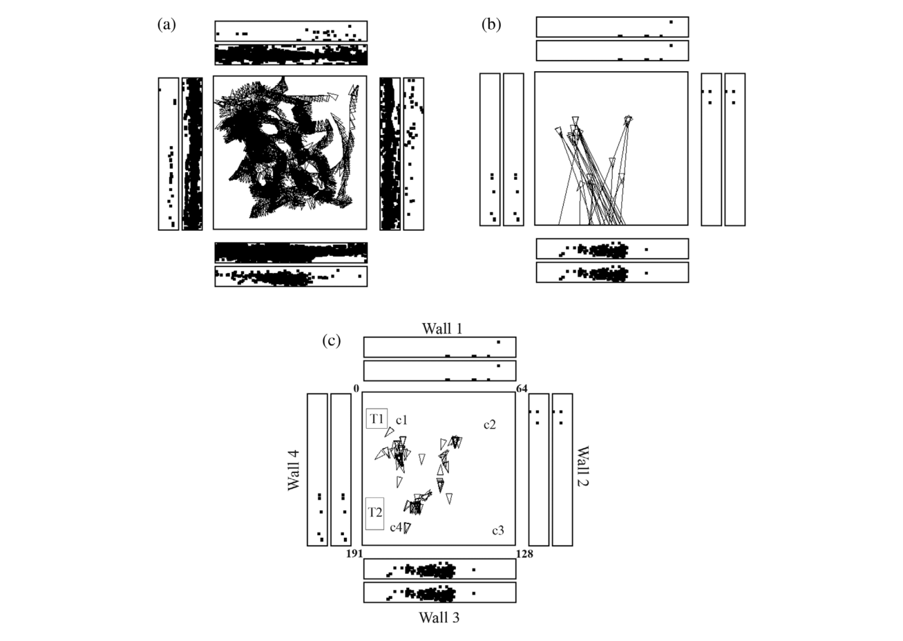
这是空间视觉细胞概念的入门图。外圈四个矩形代表房间四面墙,猴子每次放电时在其注视的墙面位置打一个点,内圈矩形标出注视点;中央方块是 2.5×2.5 m 行走区,三角标注猴子位置与头朝向。读图关键:(a) 中放电点集中在 wall 3 的一个区域——视野野在墙上而非地上;(b)(c) 显示触发放电时猴子曾处于房间里许多不同位置、头朝向各异,直接排除"place 编码"与"头朝向编码"。删除该句,或改写为'放电判据详见 Georges-François et al. (1999)'并标注非本综述内容;峰值率可引用综述给出的均值 17 spikes/s (IQR 11–20)。这张图支撑主要结果第 1 条的存在性证据。
图 2 · 空间视觉细胞 av216 的坐标不变性检验
原文图注:FIGURE 2
Testing of a hippocampal spatial view neuron (av216) to show that it has allocentric encoding, and that the response does not depend on where the monkey is located. The firing rate is shown as a function of the horizontal and vertical eye position, where positive values indicate right or up. The neuron responded when the monkey looked toward its view field (indicated with a hatched bar) relatively independently of place, eye position, or head direction. ANOVAs and information theory analyses performed on the same data cast in different ways conformed this: For spatial view, the ANOVA was p < .001 with 0.217 bits in a 500 ms period for the average Shannon mutual information; for place p = .9 with 0.001 bits; for head direction p = .5 with 0.0 bits; and for eye position p = .8 with 0.006 bits. (Modified from Georges-François et al. (1999).)
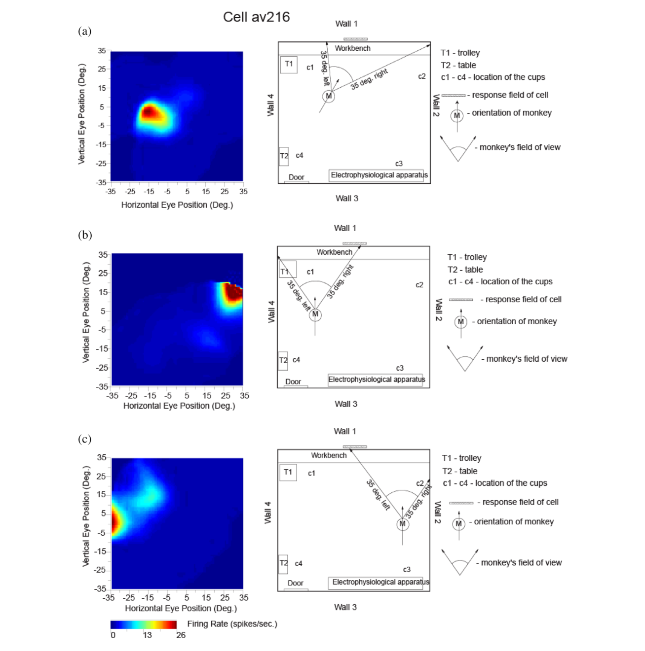
三幅子图分别对应猴子在不同位置/头朝向下以眼位坐标展示的放电率热图(正值=向右/向上)。三幅图的高放电区(阴影条标出的视野野)都落在同一空间位置 wall 1,尽管猴子所在位置和头朝向都换了——同一视野野却由不同的眼位坐标触发,这正是"世界坐标(allocentric)"的定义。附带的统计是决定性的:空间视野 ANOVA p<.001、互信息 0.217 bits;位置 p=.9(0.001 bits)、头朝向 p=.5(0 bits)、眼位 p=.8(0.006 bits)。这张图支撑主要结果第 1 条的因果排除证据,是全文最硬的单细胞数据。
图 3 · 视野遮蔽后的自身运动更新
原文图注:FIGURE 3
Self-motion (idiothetic) update of the firing of a hippocampal spatial view cell occurred for a few minutes even when the view details were obscured by floor to ceiling curtains (b). M shows the place of the monkey in the room, with the head direction indicated by the arrow. The self-motion consisted in the case illustrated by eye movements made by the monkey, but also occurred during locomotion. (Modified from Robertson et al. (1998).)
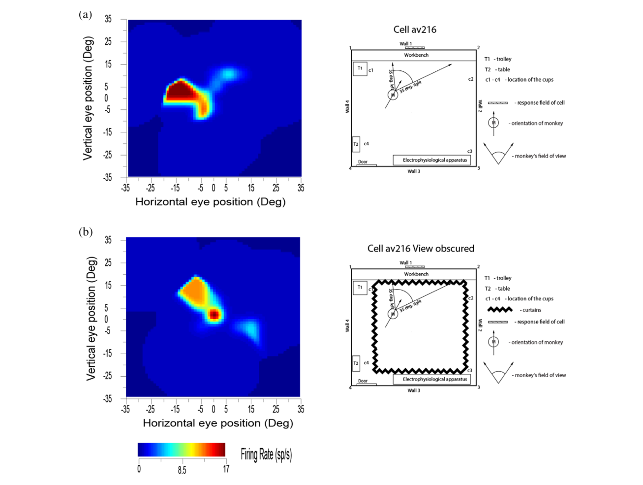
图示猴子位置 M(箭头为头朝向),(b) 为用落地窗帘完全遮蔽视野后的情形:细胞在猴子凭眼动(以及黑暗中行走)"回到"先前注视位置时仍然放电,说明注视位置表征可由自身运动信号(眼动、头动、位移的积分)更新,而不需要视觉输入;几分钟后出现漂移,也解释了为何黑暗中只能短时导航。这张图支撑主要结果第 3 条。
图 4 · 一次学习的物体—位置回忆任务
原文图注:FIGURE 4
Firing of hippocampal neurons in a one trial object-viewed location recall task. (a) In stage 1, object 1 was shown in a location on the screen being viewed, and in stage 2 object 2 was shown. In stage 3, one of the objects was shown at the top center of the screen, and the monkey had to touch the location on the screen where that object had been shown in order to obtain a juice reward. (b) A neuron that was selective for object 1 (O1) responded even in stage 4 when the object was not visible but the object and its location had to be recalled. (c) A neuron that was selective for location 1 (P1) responded even in stage 4 when the object was not visible but the object and its location on the screen being viewed had to be recalled. The average firing rate in spikes/s across trials ± SEM is shown. *p < .01; p < .05. (Modified from Rolls and Xiang (2006).)
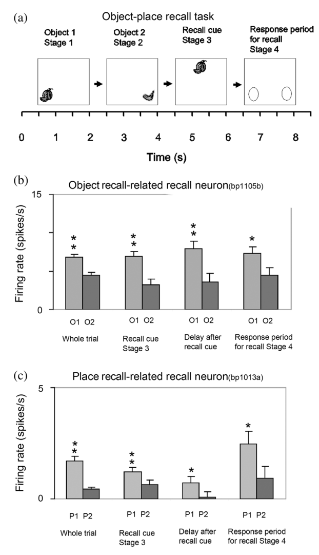
(a) 是任务时序:两个物体先后在屏幕两个位置呈现(学习,一次即学会),随后顶部中央给出回忆线索,猴子须触摸该物体先前出现的位置;(b)(c) 为直方图,横轴为任务各阶段、纵轴为平均放电率(spikes/s,±SEM,显著性 *p<.01、p<.05)。关键在第 4 阶段:线索(物体)已消失、屏幕上无刺激,对 O1 选择性的细胞和对 P1 选择性的细胞仍显著放电——放电对应的是"回忆中的物体"或"回忆中的位置"而非任何在屏刺激。这张图支撑主要结果第 4 条,是"海马实现情节记忆回溯"最直接的细胞证据。
图 5 · 人类海马系统有效连接总览
原文图注:FIGURE 5
Summary of the effective connectivity of the human hippocampal system measured across all 172 HCP participants at 7T. The maximum value of the effective connectivity is 0.2, and the strength in each direction is shown close to the termination of an arrow. The width of the lines and the size of the arrowheads reflect the strength of the effective connectivity. For areas such as the temporal lobes, the parietal cortex, and the posterior cingulate cortex, there are several subregions in the HCP atlas, and the value of the strongest effectivity connectivity to or from any subarea is shown in this case. Brain regions that are part of the ventral "what" stream are shown in blue, that are part of the dorsal "where" or "action" stream are shown in red, and that involve the orbitofrontal and anterior cingulate cortex reward value stream are in green. The ventromedial visual areas (VMV) and TH include the parahippocampal place/scene area.
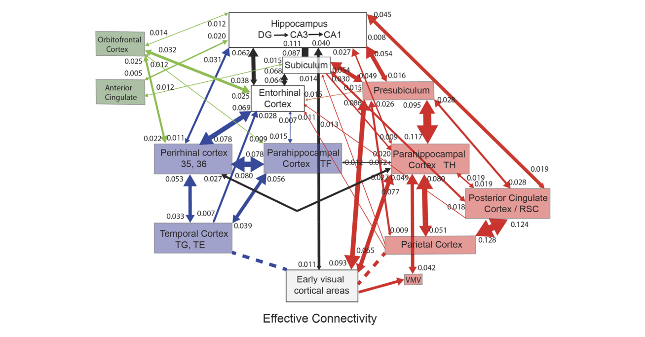
这是人类连接证据的枢纽图:172 名 HCP 被试 7T fMRI 上测得的每对脑区双向有效连接(数值标在箭头末端,最大 0.2,低于 0.010 的较强方向不显示;线宽与箭头大小反映强度)。颜色编码三股输入:蓝色为腹侧"什么"流、红色为背侧"哪里/动作"流、绿色为眶额与前扣带奖励流。读图顺序:看三条流入海马系统(海马、内嗅皮层、嗅周皮层)的通道——PHA/TH 与 VMV(含 PSA)送入场景信息,PGp 等顶叶区送入自身运动更新,眶额/前扣带送入奖励。这张图支撑主要结果第 5 条的整体架构。
图 6 · 腹内侧视觉流到场景区的连接
原文图注:FIGURE 6
Effective connectivity of the human ventromedial visual cortical stream which reaches the parahippocampal gyrus PHA1–PHA3 regions via ventromedial (VMV) and ventral visual complex (VVC) and ProStriate regions: Schematic overview. Visual scenes are represented in the anterior parts of VMV and the posterior parts of PHA1–PHA3 in what is the parahippocampal scene area (PSA; sometimes called the parahippocampal place area [PPA]) (Sulpizio et al., 2020). The retrosplenial scene area is in a band of cortex in the Prostriate cortex PRoS and dorsal visual transitional (DVT) cortex that is posterior to region RSC (Sulpizio et al., 2020). The occipital scene area is in V3CD and borders V4 (Sulpizio et al., 2020). The green arrows show how the ventromedial visual stream provides "where" inpu
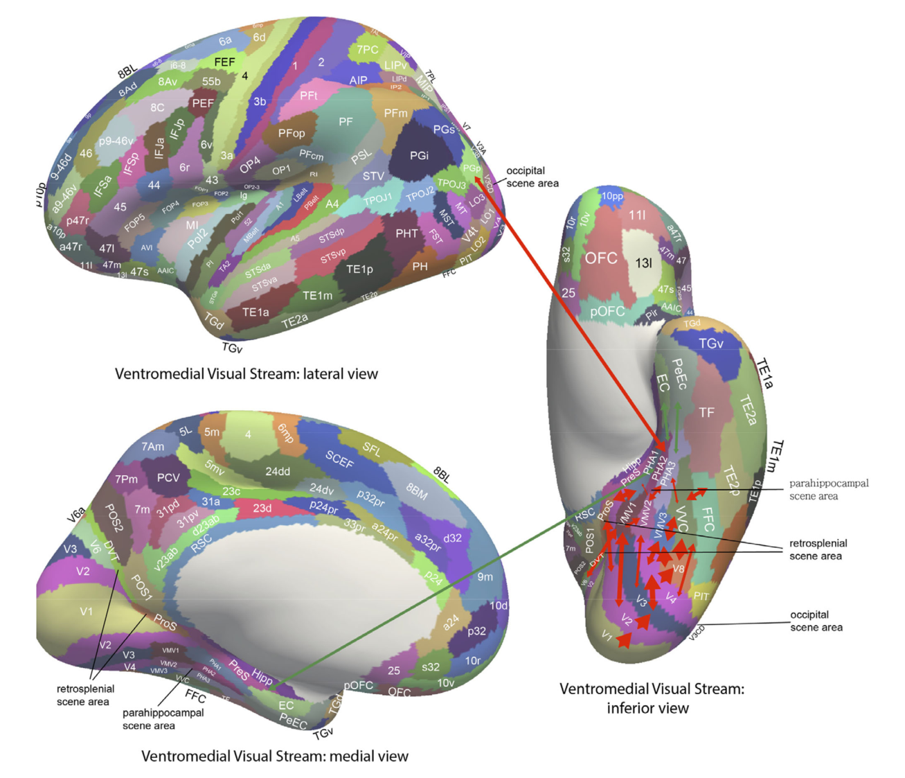
场景信息进海马的第一条通路("哪里"输入之一)。主线是 V1>V2>V3/V4→VMV1-3→PHA1-3→海马,绿色箭头标注"哪里"信息流向;PSA 位于 VMV 前部与 PHA1-3 后部交界。值得注意的细节:V2 还连到 ProS/DVT(压后场景区所在),压后与枕叶场景区(V3CD)也各有自己的场景表征带。这张图支撑"空间视觉'哪里'表征源自腹侧视觉流特征组合"这一核心假说(主要结果第 5 条)。
图 7 · 背侧视觉流与顶叶—PSA 通路
原文图注:FIGURE 7
Effective connectivity of the human dorsal visual stream which reaches (partly via V3, V3A, and LO3) the MT+ complex regions
(FST, LO1, LO2, LO3, MST, MT, PH, V3CD, and V4t), and then the intraparietal regions (AIP, LIPd, LIPv, MIP, VIP IP0, IP1, and IP2) and then the
area 7 regions: Schematic overview. Connectivity to the inferior parietal cortex region PGp, which in turn has effective connectivity to the
parahippocampal scene area in PHA1-3 is shown. Inputs to this stream from ventral stream regions such as FFC and TE2p are shown with dashed
lines. (After Rolls, Deco, et al. (2022b).)
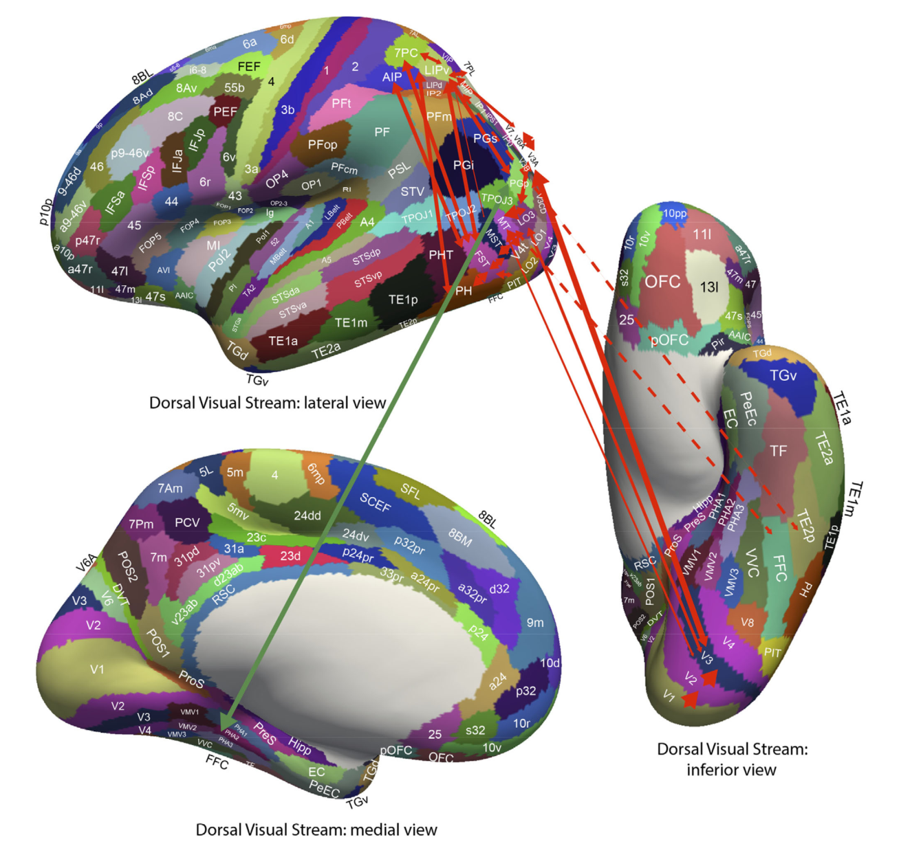
背侧流的主干:经 V3/V3A/LO3 到 MT+ 复合体诸区,再到顶内沟区(AIP、LIP、MIP、VIP 等)与 7 区。全文的关键连接是 PGp(下顶叶)→PHA1-3 中 PSA 的强有效连接——这正是把顶叶坐标变换结果(自身运动更新后的空间视野)送回场景区的通道。虚线标出腹侧流区域(FFC、TE2p)向此流的输入。这张图支撑"顶叶负责自身运动更新"的假说(主要结果第 5 条)。
图 8 · 腹外侧视觉流"什么"输入与奖励输入
原文图注:FIGURE 8
Effective connectivity of the ventrolateral visual stream which reaches inferior temporal cortex TE regions in which objects and
faces are represented (red arrows): Schematic overview. One of the red arrows shows how the ventrolateral visual stream provides "what" input
to the hippocampal memory system via parahippocampal gyrus TF to perirhinal PeEc connectivity from FFC, PH, TE1p, TE2a, and TE2p. The
green arrows show how reward regions of the orbitofrontal cortex; vmPFC (pOFC, 10r, 10v) and pregenual anterior cingulate (a24 and p32); and
punishment/non-reward regions of the lateral orbitofrontal cortex (47 m) have effective connectivity with the hippocampus (Hipp), entorhinal
cortex (EC), and perirhinal cortex (PeEC). The ventrolateral visual stream also provides input to the se
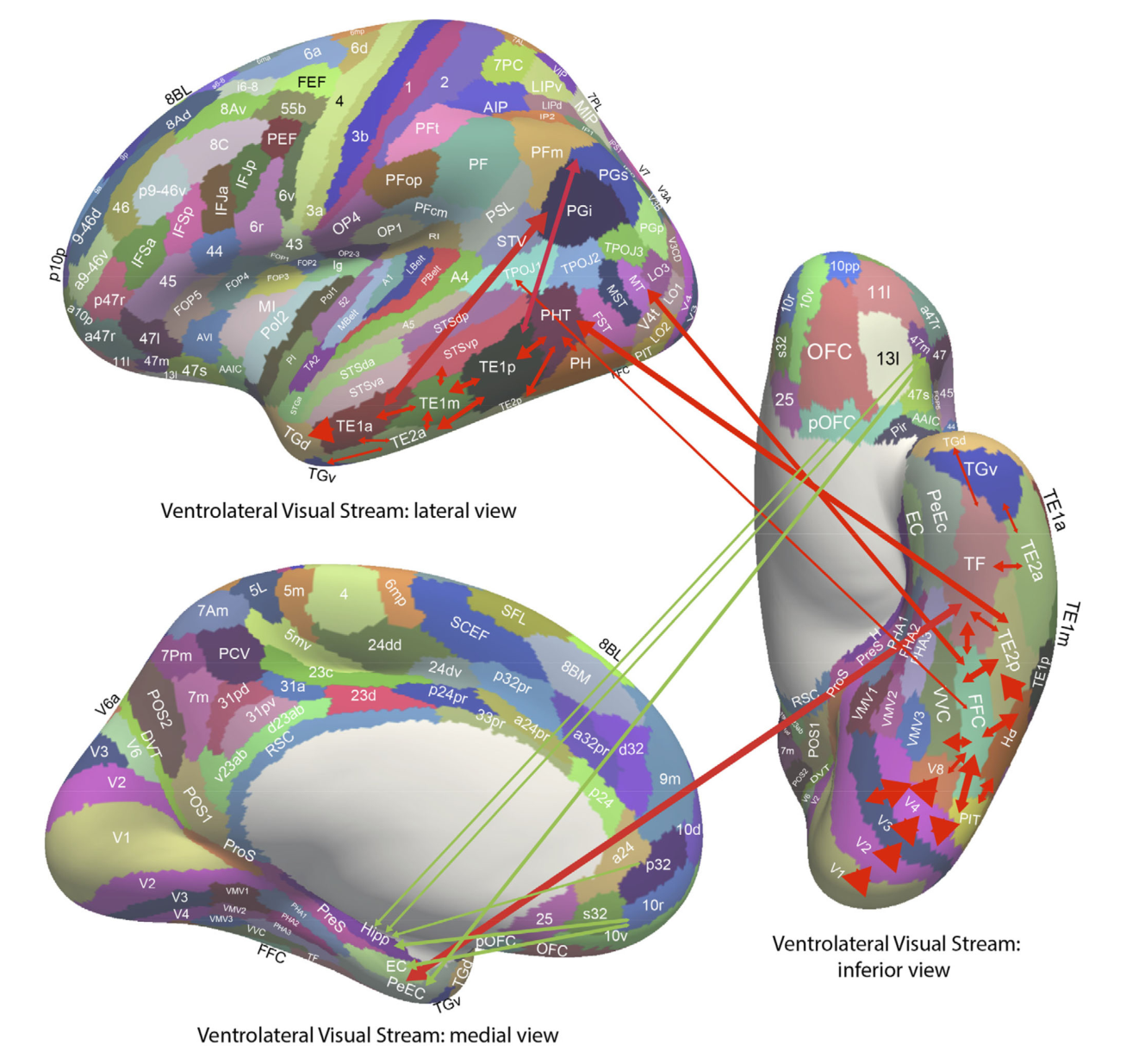
红色箭头勾勒物体/面孔的恒常表征通路(V1>V2>V4>FFC>TE),其中一支经旁海马回 TF→嗅周皮层 PeEc 把"什么"送进海马记忆系统;绿色箭头标出奖励侧:内侧眶额皮层(pOFC、10r、10v)、膝前前扣带(a24、p32)与外侧眶额皮层惩罚区(47m)连接海马、内嗅皮层与嗅周皮层。这张图支撑"what 与 reward 三路输入"假说(主要结果第 5 条),也是情绪/奖励进入情节记忆的解剖依据。
图 9 · 眶额—前扣带系统的连接综合
原文图注:FIGURE 9
Synthesis of the effective connectivity of the orbitofrontal cortex, ventromedial prefrontal cortex (vmPFC), and anterior cingulate
cortex shown in the five central ellipses, with inputs on the left and outputs on the right. The width of the lines is proportional to the effective
connectivity in the highest direction, and the size of the arrows reflects the strength of the effective connectivity in each direction. The effective
connectivities shown are for the strongest link where more than one link between regions applies for a group of brain regions. Effective
connectivities with hippocampal memory system regions are shown in green; with premotor/mid-cingulate regions in red; with inferior prefrontal
language system in blue; and in yellow to the basal forebrain nuclei of Meynert
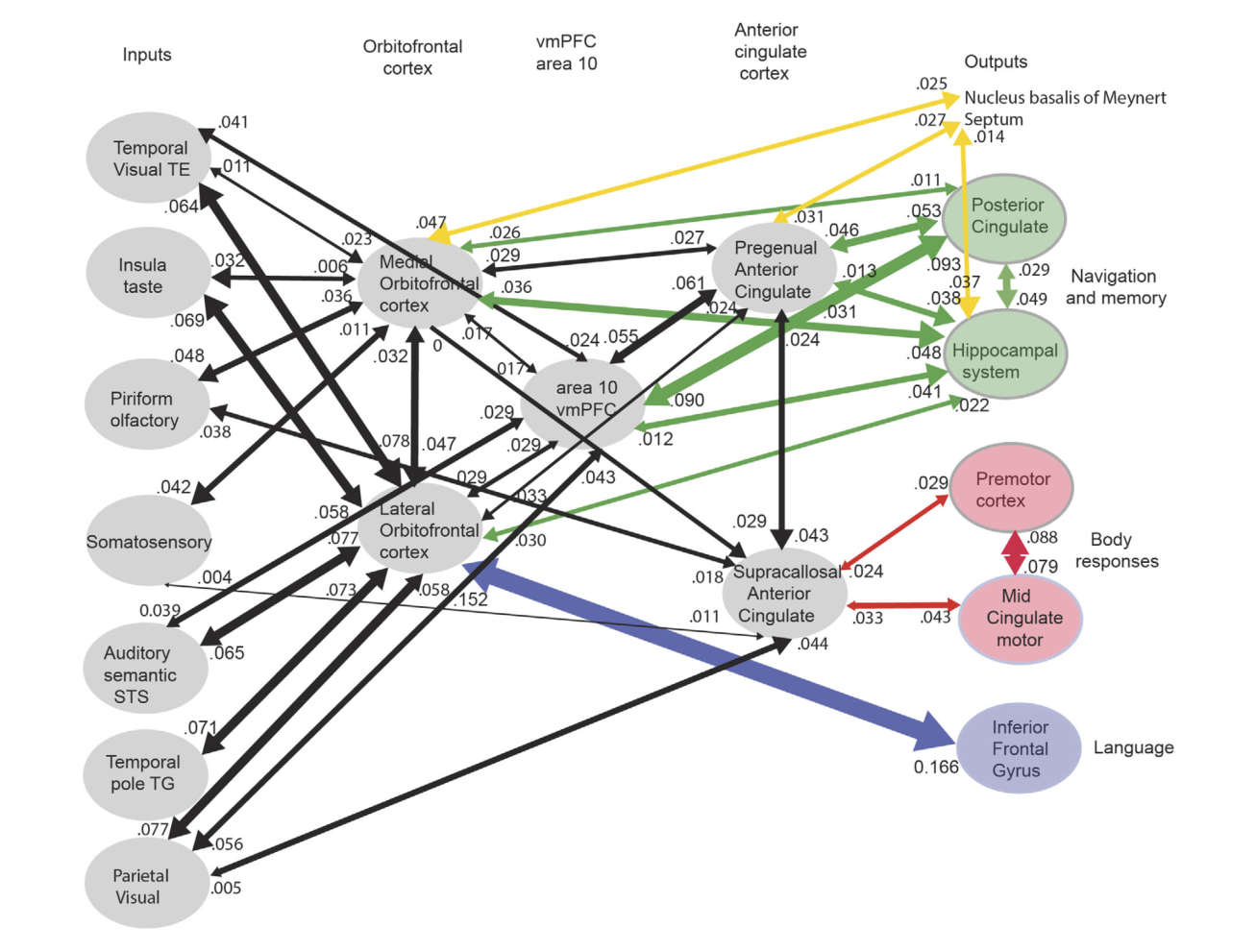
以五个中央椭圆(眶额、vmPFC、前扣带区组)为核心,左侧列感觉输入、右侧列输出。读图重点:绿色通路到海马记忆系统、黄色通路到 Meynert 基底核与隔核胆碱能神经元——这解释了奖励系统如何通过胆碱能调制影响记忆巩固(指向海马的隔核胆碱能输入与指向新皮层的基底核输入)。这张图支撑讨论中"奖励不仅进入情节记忆、还影响哪些记忆被巩固"的推论(主要结果第 5 条的延伸)。
图 10 · 海马情节记忆存储/回溯的框图
原文图注:FIGURE 10
The human/ primate hippocampus receives neocortical input connections (blue) not only from the "what" temporal lobe and
"where" medial temporal lobe scene and parietal lobe systems, but also from the "reward" prefrontal cortex areas (orbitofrontal cortex,
ventromedial prefrontal cortex [vmPFC], and anterior cingulate cortex) for episodic memory storage; and has return backprojections (green) to the
same neocortical areas for memory recall. There is great convergence via the parahippocampal gyrus, perirhinal cortex, and dentate gyrus in the
forward connections down to the single network implemented in the CA3 pyramidal cells, which have a highly developed recurrent collateral
system (red) to implement an attractor episodic memory by associating the what, where and reward component
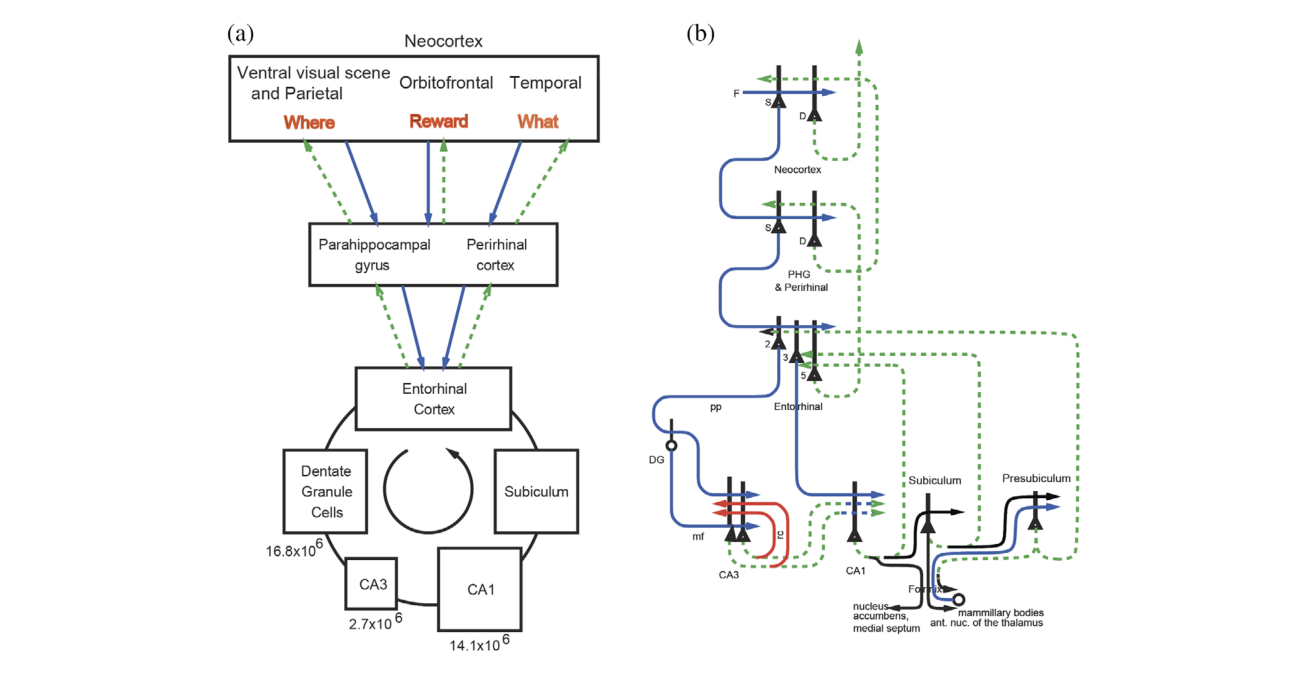
经典的 Rolls 海马架构图:(a) 框图显示蓝色前馈输入(what/where/reward 三股)逐级汇聚(旁海马回、嗅周皮层、齿状回)到 CA3,红色为 CA3 回返侧支(自联想吸引子网络),绿色为回到同一些新皮层区的回投影(用于回忆);(b) 补充主要兴奋性神经元及连接,并标注人类海马三突触环路各层神经元数量——齿状回颗粒细胞极多,支持在 CA3 之前完成"扩展编码"(expansion encoding)、生成稀疏去相关表征。这张图是理解图 12 仿真的骨架,支撑回溯理论与导航分工(主要结果第 6 条)。
图 11 · 坐标变换的增益调制模型
原文图注:FIGURE 11
(a) Gain modulation by head direction (hd) to produce a representation in allocentric bearing coordinates (relative to north) to a
location in space at which there is a landmark. The head direction is the angle between north (indicated by the long blue line) and the direction of
the head (indicated by the long black arrow). A bearing coordinate to a landmark L is represented by all combinations of head direction, eye
position (ep), and retinal position that correspond to a given bearing from the individual to a landmark in allocentric space indicated by the line
with a red arrow. The large circle is the head, and the two small circles are the eyes. The allocentric bearing to the landmark L is given by the
angle between north and the red line from the individual (observer) to the
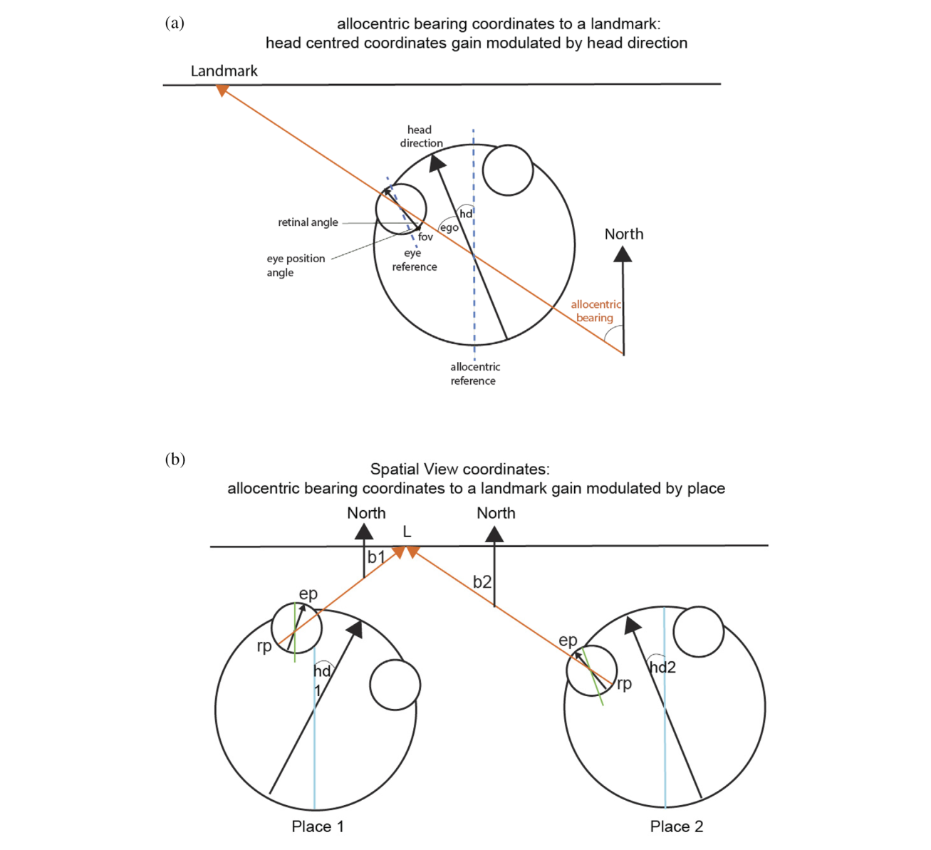
(a) 示意头朝向增益调制如何把头坐标视线角变换为对地标的"世界坐标方位角"(相对北方);(b)(图注略)再叠加位置增益调制,把对地标的方位角变换为空间视野表征——三级变换链条(眼位→头朝向→位置)正是图 7 中顶叶通路被赋予的计算功能。这张图支撑"顶叶实现自身运动更新所需坐标变换"的机制细节(主要结果第 5 条),也说明为什么导航在视野遮蔽时仍可短程维持(主要结果第 3 条的机制面)。
图 12 · 情节记忆存储与回溯的仿真
原文图注:FIGURE 12
Simulation of neocortical
"what" and "where" inputs to the
hippocampus for the storage of episodic
memory, and for the recall of "what"
(object) and "where" (spatial view)
information back to the "what" and
"where" neocortex. The pyramidal cells
bodies are shown as triangles, the
dendrites as the thick lines above the cell
bodies, and the axons as thin lines
terminated with an arrow. The
backprojection pathways for memory
recall are shown in dashed green lines,
and in red the CA3 recurrent collaterals
via which "what" and "where"
representations present at the same time
can be associated during episodic
memory storage, and via which
completion of a whole memory from a
part can occur during recall. All synapses
are associatively modifiable except for
the dentate gyrus (DG) mossy fiber (mf)
synapses on the CA3 pyramidal cells. The
dentate granule cells and the CA1 cells
operate as competitive networks
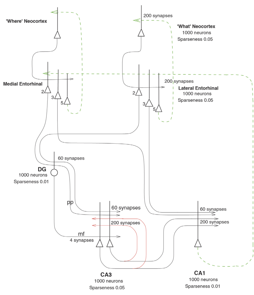
把图 10 的架构变成可运行的仿真:新皮层"what/where"输入经可修改突触存入海马,红色 CA3 回返侧支把同时呈现的 what 与 where 联结成记忆,绿色虚线回投影在回忆时把空间视野信息送回"哪里"新皮层(顶叶)。除齿状回苔藓纤维(mf,负责给每个新记忆强行分配新的一组 CA3 神经元,即模式分离)外所有突触均可联合修改;齿状回与 CA1 作为竞争网络运作。该仿真验证了从海马一路回溯到新皮层的高容量正确性,支撑回溯理论与导航分工(主要结果第 6 条)。
图 13 · 视场角决定 place cell 还是 spatial view cell 的仿真
原文图注:FIGURE 13
Simulation of rodent place cells (left) versus primate spatial view cells (right). The agent moved through a grid of all 200 × 200
places x,y. At each place, the head direction θ was rotated 5° increments. Hippocampal cells are activated by a set of three or more landmark
visual cues within the field of view of the agent α. The firing rates of the hippocampal neurons depended on the angles φ subtended by the
landmarks. The top left shows that for a rodent with a 270° field of view a combination of such cues defines a place. The top right shows that for
a primate with a 30° field of view the combination of cues defines a spatial view. The sizes of the fields of view are shown by shading. The
bottom left shows that in the simulations place fields arise with a 270° field of view, and the bottom right that spatial view fields arise on one of
the walls indicated by the rectangles when the field of view is 30°. High firing rates are indicated by yellow-red. (Details are provided in de Araujo
et al. (2001).)
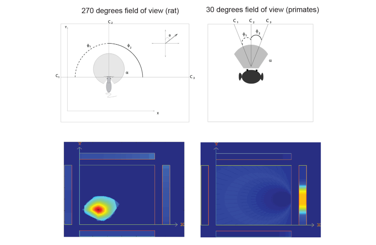
全文理论收束的一张图。仿真动物在 200×200 网格的所有地点移动、头朝向按 5°递增旋转,海马细胞由视野 α 内三个及以上地标线索的夹角 φ 组合激活。左列(270°视野)学习后生成典型 place field;右列(30°视野)生成贴在墙面上的 spatial view field(高放电率黄红色)。同样的竞争学习+自联想机制,只因中央凹有无而产出两种空间表征——这是"灵长类与啮齿类海马计算同源、表征殊异"的直接论证,支撑主要结果第 7 条。
讨论
作者的立场很鲜明:灵长类(含人类)海马的空间表征以"注视的世界坐标位置"为核心,这与中央凹视觉、眼动系统和发达颞叶的进化打包在一起;空间视觉细胞是情节记忆"哪里"成分的载体,而导航本身(坐标变换、路径整合、视觉运动动作)主要由顶叶等新皮层实现,海马只在记忆回溯参与时提供支持。人类连接证据给了这套理论三根支柱:场景输入来自腹内侧视觉流经 PSA(作者主张人类 PSA 中的神经元很可能就是空间视觉细胞——这仍是待检验的预测)、自身运动更新来自经 PGp 的顶叶通路、奖励输入来自眶额/前扣带并经胆碱能系统影响巩固。与啮齿类文献的对话贯穿全文:两者计算同源(低放电率、自身运动更新、object-place/odor-place 细胞、奖励调制、群体编码),但表征对象因视野几何而分岔;作者批评把啮齿类导航计算原样搬到人类的做法(闭眼沿路线导航的失败即可证伪其威力)。局限方面作者承认:人类海马的单细胞证据(Ekstrom、Miller、Tsitsiklis 等)大多没有同步记录眼位,"空间视野 vs. 朝向位置"的分离在人类远不如猕猴干净;虚拟环境缺乏前庭与本体感觉输入;人类 PSA 是否含空间视觉细胞是明确的未来检验点;噪声相关是否贡献信息也留待同时记录的灵长类数据。
一句话总结
这篇综述把"海马是空间记忆系统而非导航计算器"这句话写成了可检验的网络架构。依我看它最大的价值是提出了一个可以两头下注的框架:如果人类 PSA 神经元被证实就是 spatial view cells,整个"认知地图"叙事就该为灵长类改写;即使最终证明并非如此,"视野几何决定 place vs. view"的仿真本身也已把灵长类—啮齿类差异变成一个可计算的问题,而不是物种特有性的断言。
审校与证据追溯 (Verification & Evidence)
图表审计结果
- Fig1: 提取质量
good,对齐度full,识别面板[A] - Fig10: 提取质量
good,对齐度full,识别面板[] - Fig11: 提取质量
good,对齐度full,识别面板[A] - Fig12: 提取质量
good,对齐度full,识别面板[] - Fig13: 提取质量
good,对齐度full,识别面板[] - Fig2: 提取质量
good,对齐度full,识别面板[] - Fig3: 提取质量
good,对齐度full,识别面板[] - Fig4: 提取质量
good,对齐度full,识别面板[A] - Fig5: 提取质量
good,对齐度full,识别面板[] - Fig6: 提取质量
good,对齐度full,识别面板[] - Fig7: 提取质量
good,对齐度full,识别面板[] - Fig8: 提取质量
good,对齐度full,识别面板[] - Fig9: 提取质量
good,对齐度full,识别面板[]
关键事实与局限性声明
- 补充要点: Wirth et al. (2017) 星形迷宫结果的补充细节:83% 的地标反应细胞的相关特性(原文 line ~1365 'that 83% of these...')以及'许多地标反应主要出现在特定位置'这一局限讨论(line ~1259),解读未提及,该细节实际上是作者用来讨论 view 与 place 纠缠的证据之一。
- 补充要点: Mao et al. (2021) 自由移动猕猴的独立验证:22% 海马神经元表征'朝向的外界位置'而仅 5% 编码自身位置(text line 1520-1524),这是非 Rolls 实验室的近期独立证据,对'空间视觉表征是否可重复'很关键,解读完全未提。
- 补充要点: 人类连接学方法的一处限定可在解读中更明确:Figure 5 仅显示较强方向上 ≥0.010 的有效连接(图注明确说明),解读虽在图 5 注释中提到,但主要结果第 5 条引用连接强度时未带此阈值语境。